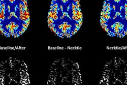
Researchers from Belgium have created virtual models of the brain individually tailored to patients' functional MR images (fMRI), according to an article published online on 28 May in eNeuro. These models can predict the effects of brain tumors and may help improve surgical planning for their removal.
When planning for brain tumor resection, clinicians typically examine functional MR images to identify key areas around the tumor as they develop a surgical strategy. But the complex dynamics of the brain make it difficult to predict postsurgical outcome based on the limited information these images provide.
One recent measure to improve the visualization of the brain has been to create more comprehensive brain models that simulate neural activity. These models can integrate fMRI data with the biophysics of the brain to predict brain function and help determine the optimal surgical approach, wrote senior author Daniele Marinazzo, PhD, and colleagues from Ghent University.
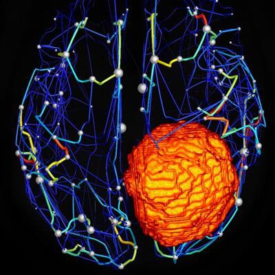
 Structural brain network of a patient with a meningioma, or brain tumor (orange). Distinct regions of the brain are depicted as spheres, with larger size indicating increasing relative importance within the network. Image courtesy of Hannelore Aerts.
To test the viability of this technique, Marinazzo and colleagues used open-source software called the Virtual Brain to construct personalized brain models. They used neuroimaging data from 25 patients who had brain tumors and who underwent an MRI exam at Ghent University Hospital between May 2015 and October 2017. They also made similar brain models of 11 patients without brain tumors.
The investigators found that studying these models allowed the surgical team to assess brain function in all of the patients, as well as accurately predict the effect of tumors on brain function. Specifically, the models revealed that the number of connections in the tumor regions of the brain was much lower (p = 0.0007) and more variable (p = 0.01) than in nontumor areas.
"Reliable prediction of patient-specific large-scale brain dynamics would open up the possibility ... to investigate what types or extent of damage the brain can withstand, and conversely, which kind of distortions can be expected after brain lesions, including those purposively induced by surgery," the authors wrote.
One of the main limitations of the study was its small sample size, but the authors plan to expand their research in the future and look into whether the virtual brain model can reliably predict postsurgical brain function.
"This [future research] would be a major step toward presurgical virtual exploration of different neurosurgical approaches and to identify an optimal surgical strategy," they wrote.