The running of the bulls at the Fiesta de San Fermín in Pamplona, Spain, occurs over a route less than 1 km, yet it's one of the most dangerous in the world. At the Spanish Society of Medical Radiology (SERAM) biennial congress last weekend, radiologist Dr. Luis Miranda Orella shared details about some of the most spectacular injuries seen at the event and also imaging's role in diagnosing casualties.
One of Spain's most iconic traditions, the running of the bulls has attracted an increasing number of participants from all over the world since it was immortalized in popular culture by Ernest Hemingway. In 2017, almost 5,500 people took part in the event, which occurs annually during the second week of July.
The run stretches over 850 meters between the Santo Domingo pens, where the bulls are kept, and the bullring, where the animals are fought to death. The run lasts less than four minutes and yet a lot of damage can happen in that time frame, Miranda told an astonished crowd of delegates at SERAM 2018.
 Above: A 25-year-old man who was stomped by a bull in the right anterior shoulder. Below A and B: Images show a right clavicle fracture with soft-tissue hematoma (arrow). C: Image shows a fracture of the right fifth rib. D: Image from a 3D scan with volume-rendering technique (VRT) shows both fractures. All images courtesy of Drs. Luis Miranda Orella and Claudio Saavedra Gutiérrez.
"Most injuries are caused by flattening, trauma, and especially bruises. Between 200 and 300 injuries are recorded every year, but only 3% are serious," he said. "Most wounds are contusions and mild injuries, and they are treated onsite in one of the 16 care stations installed along the route."
Steady rise in fatalities
Each year, an average of 45 patients are brought to the emergency department, 10 of whom require hospitalization, and 92% of all casualties are men. Only two deaths were recorded between 1947 and 1980, during the same bull run. Between 1910 and 2009, 16 fatalities were recorded, and the last three participants to die succumbed to goring (1995 and 2009) and cranial trauma (2003).
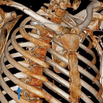
One person unexpectedly died of tuberculosis in 1910, a few months after developing an infection after receiving stitches, Miranda explained. A: A 42-year-old man gored in the right side of the chest wall. B: Coronal view shows a pulmonary contusion (arrow) and subcutaneous emphysema (arrowheads). There is no pneumothorax. C: Axial view shows the entrance wound (arrow). D: Image from 3D VRT scan shows a displaced fracture of the 10th right rib and a nondisplaced fracture of the 12th left rib.
The medical team at Navarra Hospital in Pamplona usually receives all the information on patients before they are admitted to the emergency department. Many doctors and nurses watch the spectacle on TV, so they know how patients are injured.
"Surgeons typically comment on how the bull charged the patient and already have an idea if the casualty has suffered trauma, inguinal goring, or something else. Thoracic, abdominal, and general surgeons -- and the police -- are never far away from the emergency department," he said.
Most patients admitted to the hospital present with trauma induced by falls or stamping -- either by the bulls or other participants. The most severe and spectacular injuries are caused by goring from the bullhorns, often in the left side of the thigh.
"Typically the bull and the runner run together, and either the bull touches the runner or he is trying to escape the bull. Thigh and leg injuries are by far the most common injuries, and over the past 15 years, 55 thigh and 14 leg injuries have been recorded," Miranda continued.
 A: A 49-year-old man gored five times by a bull in the right lower limb and pelvis. B: Pelvic CT image shows a fracture in the wing of the ilium (arrow) and subcutaneous emphysema (arrowheads). C: Before the CT scan, the patient had already had a first surgical management of the wound at the onsite medical facility at the bull run. Arrowheads show the direction of one of the wounds. Also, an entrance wound with surgical suture (blue arrow) and a Penrose drain (green arrow) are seen.
Modality selection
Conventional x-ray is the first-choice modality to image trauma in the limbs, but CT also is used when a case requires surgery or a second opinion. Ultrasound can be used to help detect mild abdominal trauma.
Miranda listed the four most common scenarios for trauma injuries during the running of the bulls: falls, stamping, goring, and crushing.
Injuries caused by falls are pretty straightforward to diagnose on x-ray; they typically include fractures of the head of the radius, scaphoids, and elbow luxation. Whenever necessary, x-ray images are confirmed by CT. Facial trauma also is quite common -- for example, orbital fractures and fractures of the zygomatic bone, with or without associated hematoma and emphysema.
 A: A 50-year-old woman gored by a bull in the right breast. B: CT image in the axial plane, with the arrow showing entrance wound. C: Coronal view shows subcutaneous emphysema (arrowheads). D: Axial view at a lower level shows a breast prosthesis with an irregular contour of the surface, findings suggestive of implant rupture (patient had gel effusion through the wound).
Bulls frequently run over runners, and stamping may result in a simple clavicle or rib fracture or more severe trauma, Miranda explained.
"We had a case in which five bulls had run over two men. One of them was admitted with severe trauma involving deep, important cutaneous emphysema spreading from the pelvis all the way up to the cervical vertebrae. CT showed additional rib fractures and rib fragment displacement into the thorax cavity," he said.
Goring can cause spectacular injuries, which can be undervalued at first sight.
"These injuries have an entry wound, but we don't necessarily know what happened inside the abdomen, and there can be different routes. CT is, of course, crucial in that setting," he said.
 A: Man gored by a bull. The clinical examination revealed that the wound had a superior direction. B: Coronal view shows the entrance wound above the iliac crest (arrow). Splenic laceration shows shattered spleen with active bleeding and hemoperitoneum (arrowhead). C: Image shows a fracture of the 11th left rib. D and E: Images show the extension of the hematoma and hemoperitoneum.
Miranda shared the case of a patient who presented with an injury caused by goring in his right rib. Looking at the images in detail, the radiologists noticed that the patient's right rib was pierced, and CT showed fracture on the 10th rib, in addition to mild pulmonary contusion.
"It's impressive how goring can generate such damage, with intestinal evisceration," said Miranda, highlighting the challenges of imaging such complex situations. "In that case, it was curious that the pneumoperitoneum wasn't due to the mechanical effect of goring entry, but rather to intestinal laceration, as surgery later confirmed."
Crushing is another typical scenario. In San Fermín, the most tragic crushing occurred in 1977, with one person dying of asphyxia, and again in 2013, after a door was left open too long and participants started to pour down the alley, a critical area of the route.
"That was a real catastrophe, but surprisingly few people were severely injured in the end. This could have been much worse," Miranda recalled.
One of the casualties was notably resuscitated onsite and then sent to the emergency department. CT showed that he had multiple rib fractures, pulmonary contusions, mild right pneumothorax, and pericardial effusion, probably due to cardiorespiratory arrest.