Hyperechogenic breast lesions are rare and described as generally benign. That being the case, these lesions are often overlooked and a biopsy is not recommended. But is that wise? New research suggests it is not the ideal approach.
A team, led by Dr. Eliette Helena Castillo Balladarez from Clínica Alemana in Santiago, Chile, selected biopsied hyperechogenic lesions from a database of more than 3,000 patients and found that of the 37 hyperechogenic lesions biopsied, 16 were malignant.
"Hyperechogenic cancers are very rare -- less than 2% of breast cancers -- but it is important to recognize them to avoid misdiagnosis," she told attendees at ECR 2018 in Vienna.
The history
Hyperechogenic lesions are defined as presenting greater echogenicity than subcutaneous cellular tissue of more than 90% of its volume. Stavros published a study in 1995 that gave hyperechogenic breast lesions a negative predictive value (NPV) of 100% (Radiology, July 1995, Vol. 196:1, pp. 123-134). Since then, many researchers have affirmed his results, making the notion that hyperechogenic lesions are benign a part of the collective consciousness. However, in the past few years, more researchers are questioning the finding as they've observed hyperechogenic breast cancer.
 Dr. Eliette Helena Castillo Balladarez at ECR 2018.
Castillo Balladarez and colleagues set out to evaluate the frequency of malignant hyperechogenic breast lesions, describe their imaging findings, and elucidate the histopathological characteristics. They chose cases of biopsied hyperechogenic lesions from a database of 3,394 consecutive ultrasound-guided breast core biopsies performed from 2006 to 2016, having excluded every lesion that appeared hyperechogenic due to the presence of microcalcification.
Of all the biopsies performed, 1,020 lesions (30%) were malignant. In total, 37 hyperechogenic lesions were biopsied, representing 1% of all the biopsies performed. The researchers also found the following:
- 21 benign hyperechogenic breast lesions
- 16 malignant hyperechogenic breast lesions (one ductal carcinoma in situ and 15 invasive carcinomas)
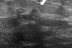
The 16 hyperechogenic breast cancers represented 1.5% of all the malignancies in the series. The women with these cancers ranged in age from 42 to 81 with a median tumor size of 26 mm. On ultrasound, three of these lesions appeared to be low-risk, while the majority of the lesions appeared to be very suspicious, especially when extrapolating the echogenicity due to margin and shapes.
Smaller tumors presented with small, central hypoechogenic foci. Larger tumors presented with a mixture of small hypoechogenic areas among extensive hyperechogenic areas.
"Another remarkable characteristic is the presence of surrounding hypoechogenic adipose tissue in almost all the lesions," Castillo Balladarez said.
Doppler ultrasound showed marked vascularization in all malignancies, demonstrating an efferent vessel even in the tiniest lesions. During histopathology analysis, the researchers found hypercellularity, fibrohyalinosis, and necrotic areas at the center of the lesions. The periphery had hypercellularity due to tumor cell proliferation, collagen filaments, and adipose tissue.
"Although the reason for the hyperechogenicity of this malignant breast lesion is still unclear, it might be explained by the presence of adipose and fibrotic tissue, multiple vessels, or, in general, due to the hypercellularity that we mentioned that creates more acoustic interfaces," Castillo Balladarez added.
The main characteristic that gives malignant breast lesions hyperechogenicity is the presence of adipose tissue mixing with tumoral cells -- most of the time it is surrounded by hypoechoic fat tissues.
It is also important to note a couple of differential diagnoses, she continued. One is pseudoangiomatous stromal hyperplasia. The other is fat necrosis.
Castillo Balladarez presented a case of a patient who came in with a left breast lesion that turned out to be fat necrosis. It disappeared within a year, but shortly after she returned with a right breast lesion that turned out to be an invasive ductal carcinoma with an appearance almost identical to the fat necrosis that she had before.
"What I want you to take home and remember is that not all hyperechogenic breast lesions are benign," she concluded. "Hyperechogenic breast cancers are rare, but they do exist. In our series, they were representative of 1.5% of all cases. It is important to recognize its ultrasound aspect and avoid misdiagnosis."