Cardiac MRI scans using a diffusion-weighted imaging (DWI) protocol have become a feasible and reproducible technique that can depict the presence of myocardial edema in ischemic and inflammatory heart disease, according to researchers from Spain who received a magna cum laude award at ECR 2018 in Vienna earlier this month.
Also, DWI can help in the characterization of cardiac masses, although there is limited experience of this area. It may be useful for follow-up and treatment monitoring in both acute myocarditis and cardiac masses, but again, there is a lack of existing data, noted lead presenter Dr. Jordi Broncano Cabrero, medical coordinator in the cardiothoracic department at Hospital Cruz Roja in Córdoba, and colleagues.
"With recent advancements in functional and molecular imaging, MRI-based sequences have made an important improvement in the characterization of normal and pathological tissues," the authors stated in their prizewinning e-poster. "DWI has become a powerful tool, not only differentiating benign versus malignant process, but also resulting in an oncological biomarker with prognostic implications in several regions of the body."
Physical basis & technique
The group's objectives were to analyze the technical adjustments required to perform DWI of the heart and to outline the real and potential clinical applications of cardiac DWI.
Ischemic heart disease constitutes the leading cause of death in developed countries, with an estimated prevalence of 8.5 million for myocardial infarction and 10.2 million for angina pectoris. It is common in people between 40 and 65 years old and is more lethal in women, they explained.
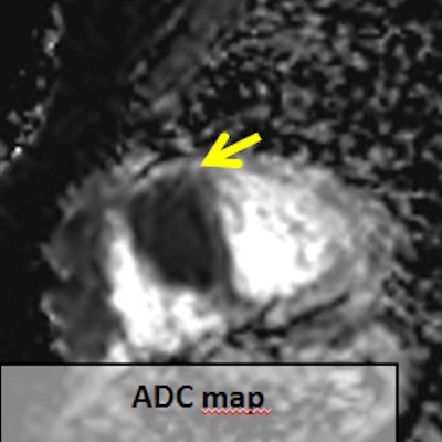
 A 60-year-old man with head and neck squamous cell carcinoma, syncope, and negative T waves in the anterior wall. During the initial examination a hypermetabolic lesion in the apical septum and apex of the right ventricle was seen (A). It shows severe restriction to DWI (apparent diffusion coefficient: 0.73 x 10⁻³ mm2/s; B, C, and D; yellow arrows). Mild hyperintensity in T2 short tau inversion recovery (F; blue arrow) and heterogeneous enhancement (E; red arrow). All images courtesy of Dr. Jordi Broncano Cabrero.
Myocardial infarction is defined as myocyte death secondary to prolonged ischemia due to overwhelmed cellular repair mechanism by inadequate blood supply to the myocardium (the ischemic cascade). Complete necrosis occurs between two and four hours after the onset of the coronary blood flow impairment.
In these patient groups, DWI involves the evaluation of Brownian motion of water molecules in biologic tissues, which has been related to tissue cellularity and architecture, the authors continued. It is based on a modified single-shot turbo spin echo sequence (Stejskal-Tanner) that focuses on the loss of signal intensity generated by means of placing several motion probe gradients surrounding a 180° pulse of radiofrequency.
For DWI acquisition, echo-planar imaging (EPI) and parallel imaging are necessary. EPI allows acquisition of the whole K space within one repetition time, while parallel imaging diminishes the time acquisition by reducing the echo train length.
"In DWI acquisition in the heart, cardiac synchronization is mandatory, usually in the diastolic phase," Broncano Cabrero and colleagues wrote. "ECG-based and respiratory navigation is very time-consuming, requiring between five to 10 minutes, depending on the number of b values included, for acquiring one slice. Breath-hold techniques usually last 15 to 25 seconds."
In other organs where there is cardiac pulsation influence in its imaging, such as the liver, free-breathing techniques are preferable over breath-hold DWI in terms of noise reduction and with subsequent increase in signal-to-noise ratio, and in their experience, free-breathing or breath-hold DWI is sufficient for assessment of the entire heart.
Three key artifacts
The following potential artifacts may hinder the performance of cardiac DWI in the evaluation of diseases of the myocardium and pericardium:
- Geometric distortion, caused by Bo inhomogeneities of the magnetic field. The solution is to increase the bandwidth and use parallel acquisition.
- Fat-saturation artifacts, due to the different processing frequency of water and fat in the phase-encoding direction. The solution is to use selective suppression techniques, such as SPIR (spectral presaturation with inversion recovery) and SPAIR (spectral attenuated inversion recovery).
- Bulk motion, caused by loss of signal intensity during high b values and motion-induced voxel misregistration. The solution is ECG triggering, breath-hold, or respiratory navigation techniques.
DWI has shown great promise in treatment monitoring and detection of recurrence of tumors. The detection of treatment response could occur as early as after one cycle of chemotherapy or one session of radiotherapy in lung cancer, and has been linked to better overall survival and progression-free survival. However, there are no reports in the literature about treatment monitoring of malignant cardiac lesions with this technique, according to the authors.
Although acute myocarditis often resolves within several weeks or months, progression of the disease to chronic myocarditis and dilated cardiomyopathy is seen in up to 21% of patients and is caused by direct viral injury and chronic immune processes. Cardiac MRI can help in the detection of persistent myocardial inflammation, precluding unnecessary endomyocardial biopsies, they concluded. Identification of edema and positive early gadolinium enhancement are independent factors for function recovery.
Editor's note: To view the authors' full range of clinical images and cases in the full e-poster from ECR 2018, click here.