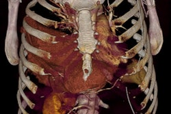
For accident victims with suspected polytrauma, a CT scan of the entire body is part of the standard of care to ensure that no injuries are overlooked. In many patients, this examination reveals secondary findings that have nothing to do with the accident, but sometimes attention is needed in the short term. For trained radiologists, it is standard practice to look at the bigger picture during an examination, thus contributing substantially to optimal care -- something brought to light during the 97th German Radiology Congress (Deutscher Röntgenkongress, RöKo 2016), which took place in Leipzig from 4 to 7 May.
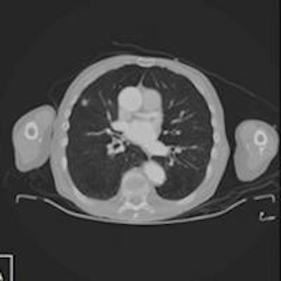
CT scans of the whole body during emergency care of accident victims are always indispensable when polytrauma is suspected. In other words, when it comes to the question of whether internal organs were damaged in addition to obvious injuries, such as in the arms or legs, CT is vital.
 Distribution of categories. Frequencies for the severity of secondary findings. The secondary findings identified during the polytrauma CT examination have been divided into four categories based on criticality:1.) Requiring immediate treatment 2.) Speedy diagnostics and/or treatment 3.) Evaluation during the symptom-free interval 4.) Normal variants or irrelevant findings. Categories: All patients, Cat 1 11%; Cat 2 13%; Cat 3 50%; Cat 4 46%. All figures courtesy of Emergency Hospital Berlin (Unfallkrankenhaus Berlin, UKB).
"The guidelines provide very clear specifications in this regard," said Dr. Thomas Kahl of the Institute of Radiology and Neuroradiology at the Emergency Hospital Berlin (Unfallkrankenhaus Berlin, UKB). He noted that all severely injured patients with high-speed trauma should undergo such an examination, as should patients with unexplained unconsciousness and victims of traffic accidents that occurred at more than 30 km/h.
Against the backdrop of these recommendations, specialist trauma centers perform full-body CT scans in patients suspected of polytrauma as a matter of daily routine. Almost six out of 10 patients present with secondary findings that require an interpretation, and nearly 800 such examinations per year are performed at the UKB alone.
 Low-grade malignant astrocytoma. Secondary finding of category 1 -- low-grade malignant astrocytoma. A and B: Axial noncontrasted CT, extensive hypodensity in right frontal region adjacent to the interhemispheric fissure. C: T1-weighted spin echo axial noncontrasted, hypodense lesion corresponding with noncontrasted CT. D and E: Fluid-attenuated inversion recovery (FLAIR), T2-weighted spin echo axial, signal alteration of the lesion. F: T1-weighted spin echo axial after gadolinium-containing contrast agent, no enrichment.
"In doing so, we keep discovering findings that have nothing to do with the trauma, but which require our attention," he said.
As part of a larger clinical study on whole-body CT, radiologists in Berlin have now taken a closer look at these secondary findings. The radiologists reported the first results during RöKo 2016. For example, of the total of 518 patients who underwent a whole-body CT scan between September 2014 and April 2015, almost 300 (57.9%) presented with secondary findings. Every 10th patient required immediate treatment.
"Among them were cases of previously undetected acute pneumonia and inflammation of the pancreas, as well as tumors of the lung and brain," Kahl said.
 Secondary findings of category 1. A and B: Polytrauma CT with coronal multiplanar reconstruction; lung lesion on the right is suspicious for malignancy. Suspicious for peripheral bronchial cancer. C and D: Polytrauma CT with coronal multiplanar reconstruction. Acute exudative pancreatitis.
In another 13.3% of patients with secondary findings, speedy diagnostics and treatment seemed advisable. Other findings involved either harmless normal variants or findings that did not require urgent attention.
To detect these secondary findings and interpret them correctly, it is essential that radiologists with broad-based training be closely involved in the clinical care of patients, he continued.
 Dr. Thomas Kahl, from Emergency Hospital Berlin.
"Instead of looking only at the bones and injured organs, we also look at the surrounding areas and go over the entire set of scans for each patient. An additional contributing factor to ensure that nothing is overlooked is that our hospital also uses the double-verification principle for polytrauma patients, with scans being independently analyzed by two different radiologists," Kahl stated.
Technological advances make it possible to keep the whole patient in view at ever-decreasing radiation doses. "The development of 64- and 128-slice CT scanners resulted in a marked drop in radiation doses in recent years," he pointed out.
But the researchers' work is not finished yet. The UKB DoReMi study, in connection with the data on secondary findings during whole-body CT scans that was collected, is currently evaluating a whole-body CT protocol that uses software optimization to achieve an additional radiation dose reduction of about 50%. The results are expected in 2017.
Editor's note: This article is an edited version of a translation of an article published in German online by the German Radiological Society (DRG, Deutsche Röntgengesellschaft). Translation by Syntacta Translation & Interpreting. To read the original article, visit the DRG website.