It's vital to understand how the anatomy affects the distribution of free gas within the peritoneal and extraperitoneal regions and use this to determine the most likely site of perforation. Identifying CT signs that allow accurate localization is also valuable, award-winning researchers from Malta and the U.K. have found.
"The presence of free gas within the abdominal cavity is an important radiological sign but has many potential causes. Some of these are life threatening and require immediate surgery, whereas some are benign and need to be recognized in order to guide conservative management," noted Dr. Kristian Micallef, a radiologist at Mater Dei Hospital, Msida, Malta.
Gas may travel from the perforation along the hepatoduodenal ligament into the fissure for ligamentum venosum and be seen adjacent to the portal vein. CT is the most sensitive imaging technique for diagnosis of free intraperitoneal air, he explained in an electronic poster that was awarded one of only five Magna Cum Laude prizes given at ECR 2013.
The key points for CT are:
Location of gas. Gas in the supramesocolic compartment, specifically gastroduodenal ligament, indicates a gastric/duodenal perforation. Gas in the inframesocolic compartment, especially in the sigmoid mesentery, indicates sigmoid perforation and is usually diverticulitis.
Secondary signs. If no obvious site of perforation is seen, look for abnormal bowel-wall thickening or mesenteric stranding.
Site of perforation. Occasionally, a defect will be seen in the bowel wall, indicating the exact site of perforation.
"When an abnormal intra-abdominal gas pattern is detected, it must be carefully analyzed. Is the gas within a loop of bowel? Multiplanar reconstructions can help to resolve this. Also, has the patient had recent surgery or instrumentation that could explain the presence of gas? Is the patient acutely unwell? If not, the gas could represent a benign cause of pneumatosis intestinalis," Micallef stated. "Knowledge of the anatomy of this region and specific imaging findings can guide the radiologist to the correct diagnosis in the majority of patients."
Intraperitoneal organs invaginate and are almost completely covered by the visceral layer. Extra- or retroperitoneal organs lie outside the peritoneal cavity, either exterior or posterior to the parietal peritoneum. These organs are usually only partially covered by peritoneum, he explained.
The embryological development of the abdominal cavity determines the formation of the communicating peritoneal spaces. In the developing fetus, the peritoneal cavity is divided into right and left by the ventral and dorsal mesenteries of the primitive gut. Many of the ligaments and mesenteries are formed from remnants of the ventral and dorsal mesenteries.
"The detection of intra-abdominal gas on imaging raises the possibility of multiple diagnoses. Often, it is the result of perforation of a viscus, most commonly a gastric/duodenal ulcer or perforated diverticulitis," he emphasized. "Other causes include a perforated malignancy of the gastrointestinal tract and abdominal trauma. Benign causes such as recent instrumentation or pneumatosis intestinalis should also be considered."
Radiologists must try to locate the site of perforation because this will allow the surgeon to determine whether or not surgery is indicated and to plan the site of incision, which is especially important if laparoscopic surgery is planned, according to Micallef and his co-authors from London.
The peritoneum consists of two continuous layers: the parietal peritoneum lines and attaches, in places, to the internal surface of the abdomino-pelvic cavity, while the visceral peritoneum invests and covers the external surface of viscera. Remnants of the dorsal mesentery form the gastrocolic, gastrosplenic, gastrophrenic; gastropancreatic ligaments form part of the greater omentum; and the splenorenal and phrenicocolic ligaments also arise from the dorsal mesentery.
The supramesolic compartment is divided into left and right by the falciform ligament and it contains the stomach, liver, and spleen, they wrote. The ventral mesenteric remnant gives rise to the falciform ligament, the gastrohepatic and hepatoduodenal ligament, and the latter two form part of the lesser omentum.
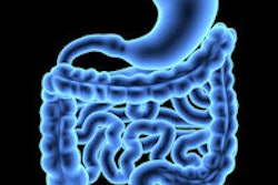
Under normal circumstances, no gas should be identified within the peritoneal or extraperitoneal spaces, but if the gastrointestinal (GI) tract is damaged -- most commonly by inflammation, malignancy, or trauma -- gas will leak from the GI tract into the surrounding peritoneal or extraperitoneal spaces. Unlike free fluid, gas will rise to lie against the anterior abdominal wall in the supine patient. The site of perforation and the peritoneal connections it can travel along will determine the location of the gas, the authors stated.
"Perforation of the GI tract does not always result in free gas; sometimes surrounding inflammatory change will wall off the abnormal segment, resulting in a fluid collection. This is commonly seen in appendicitis and diverticulitis," they elaborated. "When a patient presents with suspected perforation, clinicians will usually request a supine abdominal radiograph and erect chest radiograph. These may demonstrate free gas, but sensitivity and specificity are low, the site of perforation cannot be determined, and the images are difficult to interpret. Normal radiographs do not exclude the diagnosis."
CT should be the first-line test for most patients with an acute abdomen, they continued. In a patient with an acute abdomen with suspected perforation, a portal venous phase postcontrast scan should be obtained through the abdomen and pelvis, and oral contrast is not required. The scan must be carefully assessed using multiplanar reconstructions and altering the windows to increase conspicuity of small gas bubbles. Care must be taken to follow bowel loops to establish whether any gas seen is within the bowel lumen and the bowel wall. An accurate diagnosis of the site of perforation is feasible in 86% of cases, they maintained.
Perforation of a peptic ulcer is the most common cause of pneumoperitoneum. Anterior wall ulcers of the stomach and duodenal bulb usually perforate freely into the intraperitoneal space, initially supramesocolic, whereas posterior wall gastric ulcers perforate into the lesser sac. Some ulcers will seal off immediately, and the only radiological sign of perforation may be gas adjacent to the stomach or in the gastroduodenal ligament, the authors concluded.


