PACS technology, combined with Web-enabled viewing, often is praised for making a patient's diagnostic images rapidly available to any authorized clinician who needs to see them, but this access can be at the expense of face-to-face discussions between radiologists and their colleagues. At the Norwegian Radium Hospital in Oslo, PACS plays a major role in improving face-to-face communication.
The 400-bed Norwegian Radium Hospital is a national center for cancer treatment in Norway, and staff members conduct research on an international scale at the Institute for Cancer Research. These institutions, affiliated with Oslo University Hospital, offer a useful combination of patient treatment and research in the Scandinavian region.
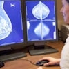
 Staff in the radiology department at the Norwegian Radium Hospital take part in multidisciplinary weekly meetings to discuss difficult cases. All images courtesy of Agfa HealthCare.
Radiology plays a major role at this hospital. More than six terabytes of image data are archived each year. Approximately 80,000 images are acquired each day. The average study contains more than 460 images; some studies have thousands of images. This is not unusual, considering that two-thirds of the exams performed in the radiology department are CT exams. To add to the volume, cancer patients throughout Norway who are referred to the hospital bring their prior digital studies on CD or DVD for inclusion into their electronic records.
Managing all these images would not be possible without an integrated radiology information system and a PACS, which the hospital began to use in 2003. In 2009, additional clinical application modules were added that were intended to improve the display and workflow management of high volume studies, as well to help make diagnostic interpretation easier for the radiologists.
The center's radiologists, radiation oncologists, and cancer treatment teams attribute the advanced clinical applications of their PACS (Agfa HealthCare) for making their meetings about patient cases much more comprehensive and valuable, not to mention more efficient.
The display and image manipulation capabilities of IMPAX Volume Viewing are not unique to the Agfa PACS. Most high-performance PACS offer their functionality. They help radiologists save time while interpreting studies, especially at cancer research centers. Many research protocols are more image-intensive than routine clinical imaging, and they can also require more types of imaging using different modalities.
Agfa offers several volume-viewing modules to supplement the core functionality of its diagnostic workstation software. These are used for reformatting cross-sectional imaging, 3D volume rendering and segmentation, vessel viewing, PET/CT viewing, and sophisticated comparison of series of images from two different exams.
At Norwegian Radium Hospital, the functionality of the volume viewing modules has dramatically improved the way findings of a patient's images are conveyed and discussed. Multiple daily meetings are held to discuss the cases of patients who are scheduled for surgery or radiotherapy treatment. The radiology department also hosts multidisciplinary team meetings that are held on a weekly basis.
"Our mornings are filled with meetings," said Dr. Cathrine Saxhaug, a specialist in oncologic radiology. These daily meetings tend to be organized by cancer type, such as a meeting about patients with brain tumors. "We typically spend between 10 and 30 minutes to show and discuss the latest radiological findings of seven to eight patients with their oncologists, the medical physicists who plan radiotherapy treatments, and the radiation therapists who perform it."
The multidisciplinary weekly meetings are used to discuss difficult cases. Many more types of specialists attend: oncologists, surgeons, pathologists, gynecologists, nurses, and medical physicists. These tend to be problem solving and treatment planning meetings, taking between one to two hours.
A tight integration with the PACS makes volume viewing highly efficient by making the loading of images seem seamless. Workstation images are projected on large screens, one showing the PACS "control" workstation screen and two others showing volume-viewing displays.
"The volume-viewing functionality of the PACS makes it easier to display large volume datasets in ways that make sense to everyone. We use it as a tool to demonstrate findings in a better way. We do a lot of 3D volume imaging, so we need to have a system that displays the volumes effectively."
 The modern design of the 400-bed Norwegian Radium Hospital contributes to the high-tech feel of the center.
"Better visualization of image volumes makes it easier to initially interpret the images and to find pathologies. We can show precisely where the pathology is compared to all the neighboring structures. Colorized subtraction images can highlight changes in a patient's anatomy. Segmentation tools allow bone and other obscuring structures to be hidden," she explained.
"The ability to compare two sets of images effectively by using a registration tool is an efficient way to monitor tumor development and response by the tumor to treatment over time," she said. "To display the growth of a brain tumor, for example, we use the 3D volume-viewing feature where both new and old exams are displayed, automatically segmented, and fused.
And if they want to retain specific displays, including 3D and image animation, or to highlight a finding in different orientations, all of this can be retained as a file for future review and sent to the PACS. Surgeons and oncologists do not use the volume-viewing tools themselves, but they use what the radiologists create for them, even taking these images into operating rooms and treatment rooms.
Saxhaug and her colleagues, as well as radiologists who practice in cancer centers worldwide, struggle each day with the best ways to manage the steady increase of images and data along with their complexity. They want to make these images and data relevant to their colleagues. All work fast-paced days.
Advanced functionalities of PACS, when used as a communication tool as it is at the Norwegian Radium Hospital, can help.