British researchers are using MRI to image hemorrhaging inside the heart to determine how much damage has been caused by a heart attack.
In a study published online in Radiology (January 21, 2009), researchers from the Medical Research Council Clinical Sciences Centre in London, led by Declan O'Regan, Ph.D., assessed the feasibility of using multiecho T2-weighted mapping to detect myocardial hemorrhage after percutaneous primary coronary intervention for acute myocardial infarction.
The study enrolled 13 men and two women with a mean age of 59 years, ranging from 41 to 74 years. All 15 patients had undergone percutaneous primary coronary intervention within the previous seven days.
The study criteria included men and women who had an electrocardiographic diagnosis of myocardial infarction and coronary intervention within 10 hours of the start of symptoms. All patients presented within 24 hours of the onset of chest pain for treatment of a first myocardial infarction.
Study protocol
Cardiac MRI examinations were performed with a 1.5-tesla scanner (Achieva, Philips Healthcare, Andover, MA), with a five-element cardiac phased-array coil. An intravenous bolus of gadolinium dimeglumine (Magnevist, Bayer Schering Pharma, Berlin) was administered at a dose of 0.2 mmol/kg of body weight.
Two observers, each with five years of experience in cardiac MRI, performed all cardiac MRI measurements independently. Initially, the images were masked to include only the myocardium for analysis.
The study noted that each observer "defined a point at the edge of the hyperintense zone and a boundary was then automatically generated around the [ischemic area of risk (IAR)]. This method was compared with a conventional signal intensity threshold level method by using two, three, and five standard deviations (SDs) above the mean of remote normal myocardium."
Study results
Cardiac MRI was performed one to seven days (mean, 3.2 days) after percutaneous primary coronary intervention. Physicians intervened in the left anterior descending artery in seven patients, the right coronary artery in six cases, the circumflex artery for one patient, and the obtuse marginal artery in one case.
 |
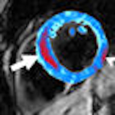
| Image of a left anterior descending artery occlusion acquired by T2-weighted MRI prior to contrast administration. Red area shows a region of postreperfusion hemorrhage (arrow). Susceptibility artifact adjacent to inferolateral epicardium was manually excluded (arrowhead). Image courtesy of the Medical Research Council Clinical Sciences Centre in London and Radiology. |
"Transmural edema was present in all subjects on the T2-weighted short inversion time inversion recovery images and was in the same anatomic region as the late gadolinium enhancement," the authors wrote.
The mean area of hemorrhage, as indicated by a T2 measurement of less than 20 msec, was 5% at the level of the infarct. The researchers set the 5% value of the hemorrhage area as the cutoff value for subsequent subgroup analysis. Six of the 15 patients demonstrated 5% or more hemorrhage after reperfusion.
By using a tone detection method, the mean ischemic area of risk in all subjects was 34% (range, 20%-57%). The researchers found that the mean ischemic area of risk was larger in subjects with 5% or more hemorrhage with a mean size of 44%, compared with 27% in subjects with less than 5% hemorrhage.
Area of risk
With an accurate measurement of the ischemic area of risk, the researchers noted that physicians can improve myocardial salvage.
One limitation cited by the study authors was that the threshold level for detecting hemorrhage at cardiac MR was not determined on the basis of histologic data, as none of the prospectively recruited patients died. "However," they wrote, "there are autopsy case reports demonstrating close histopathologic correlation of the hypointense T2 signal with postreperfusion hemorrhage in human and animal studies that show that T2 and T2 sequences accurately localize blood products."
The authors recommended that studies using cardiac MRI to "determine the IAR and myocardial salvage should use boundary detection methods for quantification, as arbitrary signal intensity threshold levels are unreliable when hemorrhage is present."
Postreperfusion hemorrhage, they added, can be assessed by using T2 mapping and may provide an imaging marker of poor myocardial salvage.
By Wayne Forrest
AuntMinnie.com staff writer
February 13, 2009
Related Reading
CMS launches national coverage analysis of cardiac MR flow studies, January 22, 2009
MRI can provide safe imaging for pediatric heart patients, August 29, 2008
MRI evaluation of chest pain cuts acute coronary syndrome, August 15, 2008
MRI edges out CT in postinfarct myocardial studies, May 8, 2008
MR studies gauge heart risk after myocardial infarction, October 15, 2007
Copyright © 2009 AuntMinnie.com