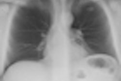
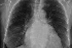
A study by Italian researchers has added to the growing body of evidence indicating that digital radiography (DR) with a tomosynthesis capability is superior to conventional DR in detecting pulmonary alterations that could be signs of lung pathology. But tomosynthesis wasn't totally without drawbacks.
A research team led by Dr. Emilio Quaia from the University of Trieste described their preliminary clinical experience comparing a digital tomo unit to conventional DR in diagnosing lung parenchyma alterations. They presented their results at the 2008 RSNA meeting.
Digital tomosynthesis units began shipping commercially in the U.S. in 2007; when clinical research studies began appearing in 2008, they found that the units offer dramatically improved sensitivity compared to conventional DR.
Vikgren and colleagues found that digital tomosynthesis had a sensitivity of 92% for lung lesions versus 28% for standard DR in a study published in Radiology in October 2008, while Dobbins et al reported a sensitivity of 70% for digital tomo against 22% for conventional DR in a paper in Medical Physics in June 2008.
Quaia and colleagues chose to focus on suspected alterations to pulmonary parenchyma that could indicate the presence of lung cancer. "Since lung cancer is the most important alteration of the lung parenchyma, the principal aim was to exclude the presence of lung cancer," Quaia said in an e-mail to AuntMinnie.com. Patients thus excluded would have no need to be worked up with CT, reducing radiation dose and saving healthcare costs, he added.
The prospective study focused on alterations ≤ 4 cm in a patient population of 37 subjects, who had a total of 60 pulmonary alterations. The study cohort consisted of 24 men and 13 women, with a mean age of 72.7 years (± 7.9 years).
In addition to conventional DR (Definium 8000, GE Healthcare, Chalfont St. Giles, U.K.), patients underwent digital tomosynthesis with a commercially available system (VolumeRad option for Definium 8000). Patients were also scanned by 64-detector-row CT to confirm the presence of lung alterations.
Technical parameters used for digital tomo imaging were a tube energy of 125 kVp, with a 0.6-mm nominal focal spot and additional 0.1-mm copper beam filtration. The tomosynthesis unit collected an average of 53 projection images in a 35° arc around the patient, with an interval between slices of 5 mm. Total acquisition time was 12 seconds.
Images were reviewed by two readers, one with three years of experience and one with 20 years. Observer 1 found 57% of lesions (34 of 60) with conventional DR, a number that jumped to 90% (54 of 60) with tomosynthesis. Observer 2 found 62% of lesions (37 of 60) with DR, a figure that jumped to 93% (56 of 60) with tomosynthesis.
In particular, the group found that digital tomosynthesis expanded radiography's detection limit to substantially smaller nodules -- those greater than 4 mm -- than previously possible. Such nodules are within the criteria endorsed by the Fleischner Society as being appropriate for additional workup, Quaia said.
"Considering only the nodules that would be actionable, in our study more than 90% were visible in the tomosynthesis images compared to less than 70% in the digital conventional radiographs," Quaia told AuntMinnie.com. "Consequently, digital tomosynthesis provides reliable and immediate detection of nodular lesions which deserve a further analysis by chest CT."
There was a price for this increased diagnostic performance, however: radiation dose. The digital tomosynthesis technique produced an average patient dose of 0.2 mSv per exam, more than triple the 0.06 mSv for a standard posteroanterior/lateral DR chest sequence.
Quaia and colleagues pointed out, however, that digital tomosynthesis' dose compares to 1-8 mSv for a CT study of the same anatomical region, which currently occurs because tomo is not part of the diagnostic workup for suspicious lesions.
Another limitation is tissue blurring that occurs outside the region of interest due to the filtered back projection image reconstruction algorithm that the group used, which Quaia described as "imperfect." The group is working on other reconstruction algorithms, he said.
In their conclusion, the researchers said that digital tomosynthesis could become a useful tool in the workup of suspicious pulmonary lesions. In future research, the group plans to increase the patient size in their studies of lung alterations, and they also intend to examine the use of digital tomosynthesis in an emergency skeletal x-ray unit to improve fracture detectability.
By Brian Casey
AuntMinnie.com staff writer
February 18, 2009
Related Reading
Digital chest tomo beats standard DR for pulmonary nodules, October 21, 2008
Digital tomosynthesis beats standard DR in finding chest lesions, August 11, 2008
TMJ imaging benefits from digital tomo technique, December 24, 2007
DR pumps new life into tomosynthesis-based radiography, October 29, 2007
Duke researchers combine DR tomosynthesis with 3D CAD, June 4, 2007
Copyright © 2009 AuntMinnie.com