Italian researchers recommend coronary MR angiography (MRA) as the modality of choice in the follow-up of patients with Kawasaki disease to reduce the potential cumulative effect of radiation exposure from repeat CT angiography (CTA) exams.
The use of CTA should be restricted to cases when physicians cannot obtain diagnostic images from MRA, added the researchers from the University of Rome "La Sapienza."
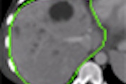
Kawasaki disease is an acute systemic vasculitis that generally affects children younger than 3 years old and can lead to coronary artery disease and stenosis in as many as 25% of afflicted patients. The disease also can cause aneurysms in coronary arteries, which may rupture or lead to thrombosis and/or complete occlusion.
"That is why patients with Kawasaki disease need coronary artery imaging for risk stratification and therapeutic management as follow-up throughout their lives," said lead author Dr. Emanuela Algeri.
Modality of choice
Although selective coronary angiography currently is the imaging modality of choice for these patients, MDCT angiography in the past few years has proved to be a valuable diagnostic method as well. However, selective coronary angiography is an invasive method and both selective coronary angiography and CTA use ionizing radiation.
"We should consider the estimated risk of radiation-induced cancers in children, especially if they need to undergo many repeat exams throughout their lives," Algeri said while presenting the findings at the American Roentgen Ray Society (ARRS) annual meeting.
For their study, the researchers enrolled 15 consecutive patients with a mean age of 17.6 years (11 males, four females) and a previous diagnosis of Kawasaki disease. The patients underwent both MRA and CTA, with those results compared to previously performed selective coronary angiography.
MRA was performed using a 1.5-tesla system. The protocol included free-breathing acquisition with no contrast. After injection of a blood-pool contrast agent, physicians performed a breath-hold MRA acquisition, focusing on the vessel of interest.
CTA protocol
CTA was performed on a 64-slice CT scanner after the intravenous injection of a 70 mL bolus of a high-iodine concentration nonionic contrast agent (Iomeron [iomeprol], Bracco Diagnostics, Milan, Italy). To reduce radiation dose, the researchers used both electrocardiogram pulsing and an automatic exposure control system.
Results of the two imaging techniques were compared with findings from previously performed selective coronary angiography.
The researchers found that coronary CTA produced diagnostic images in all 15 patients, while coronary MRA produced diagnostic images in 13 of 15 patients. All CTA findings matched the previously verified selective coronary angiography images.
"We could identify four small aneurysms, 15 medium-caliber aneurysms, 13 giant aneurysms, three segments with stenosis, and nine segments with occlusion," Algeri said. Six of nine segments with occlusion were within an aneurysm.
MRA 'correlated well'
The 13 diagnostic-quality exams provided by MRA also "correlated well with selective coronary angiography and CT angiography findings," according to the researchers. "In particular, the relation between the caliber of aneurysms measured by MR angiography, 64-slice CT angiography, and selective coronary angiography were high," Algeri added. "However, stenosis grading was much more difficult."
MRA is a useful diagnostic tool for the follow-up of patients with Kawasaki disease, the researchers concluded. "We should consider [MRA] as the first technique to follow up patients," Algeri said. "CT angiography should be recommended only when we cannot obtain diagnostic images with MR."
The researchers also noted some disadvantages with MRA. While the major advantage of MRA is the lack of ionizing radiation, the imaging modality's spatial resolution "is lower than that of coronary CT angiography." Algeri said. "This could be a problem when evaluating distal branches and stenosis grading."
An advantage of MRA is the ability to perform perfusion evaluation and viability studies in the same scanning session. However, MRA "has a long scan time compared to CT angiography," she added, "even if [MRA] doesn't require breath-holding."
By Wayne Forrest
AuntMinnie.com staff writer
July 6, 2009
Related Reading
MR angiography planning halves radiation, contrast dose during UAE, May 28, 2009
Quantitative MR angiography shows promise in stenosis detection, February 12, 2009
AHA urges cautious use of coronary CT angiography and MR angiography, July 9, 2008
Studies outline techniques for ramping up MR in renal artery imaging, January 10, 2008
MR angiography offers greater detail in vascular imaging, March 10, 2006
Copyright © 2009 AuntMinnie.com