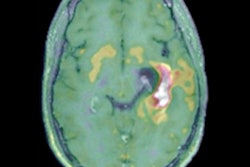
An artificial intelligence (AI) algorithm can identify sarcopenia on routine brain MRI scans of patients with glioblastoma, enabling predictions of how long the patient might survive this aggressive cancer, researchers are reporting at this week's National Cancer Research Institute (NCRI) Virtual Showcase in the U.K.
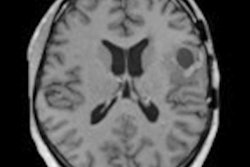
A team of researchers led by Dr. Ella Mi of the Imperial College London trained a deep-learning algorithm to quantify the temporalis muscle on routine brain MRI scans performed on glioblastoma patients. They found that the AI model's measurement of the temporalis muscle's cross-sectional area (CSA) prior to treatment was a significant predictor of both overall and progression-free survival.
"This has the potential to improve prognostic estimates and could be used to plan treatments," said Mi in a statement issued ahead of the event by the NCRI. "For instance, previous evidence has shown that frail patients might benefit from shorter courses of radiotherapy, or chemotherapy with temozolomide alone. It could also guide therapeutic interventions for muscle preservation, including nutritional support, exercise therapy, and drugs."
After being diagnosed with glioblastoma, a patient has an average of 12-18 months to live, and less than 5% are still alive after five years. As some patients do better than others, objective assessments of a patient's frailty and physical condition are needed in order to guide decisions on treatment, diet, and exercise, according to the researchers.
In the hope of developing such an objective measurement, the researchers trained a convolutional neural network to quantify the cross-sectional area of the thickest part of the temporalis muscle on brain MRI scans acquired during diagnosis and follow-up care.
"We realized that sarcopenia could be identified by quantifying muscle in cross-sectional imaging that cancer patients routinely undergo," Mi said. "This would allow for opportunistic screening of sarcopenia as part of cancer care without additional scanning time, radiation dose, or cost."
The study, which involved 152 brain MRI scans of 45 patients from the Imperial Tissue Bank and the Diffusion in Glioma Study, found that these measurements were associated with both overall and progression-free survival.
| Average length of survival in glioblastoma patients | ||
| Patients with low temporalis muscle cross-sectional area measured by AI | Patients with high temporalis cross-sectional area measured by AI | |
| Average length of survival | 14 months | 21.3 months |
What's more, even after taking into account factors such as age, sex, the side of the brain the tumor was on, and a genetic characteristic of brain tumors that often predicts chemotherapy response, the researchers found that patients with high CSA results had approximately 60% lower risk of death and 75% lower risk of disease progression compared with patients who had low CSA measurements.
"We are the first to show that this measurement of sarcopenia, generated automatically from routine imaging is sufficiently accurate and reliable to be a useful prognostic marker in cancer, while taking substantially less time than trained humans," Mi said.
The researchers are now validating their results on a different set of patients, as well as performing additional studies to characterize changes in the temporalis muscle during follow-up of glioblastoma patients after diagnosis and treatment. In addition, they said they would also like to perform a multicenter study by collaborating with other academic centers that have repositories of glioblastoma MRI exams.