Award-winning German researchers have developed a clinically viable method for imaging of the extracranial arteries in cases of acute ischemic stroke without the use of gadolinium contrast agents.
"There is great demand for noncontrast-enhanced MR angiography (CE-MRA) techniques providing high image quality in a short scan time," noted Dr. Lenhard Pennig, from the Institute for Diagnostic and Interventional Radiology, Faculty of Medicine, and University Hospital in Cologne, Germany, and colleagues in an ECR 2020 e-poster that received a cum laude award from the judges.
"3D relaxation-enhanced angiography without contrast and triggering (REACT), a combination of T2 preparation and non-volume-selective short tau inversion recovery pulses with mDIXON XD readout, was introduced. It combines benefits of steady-state free precession such as bright-blood signal with robust suppression of fat and background for flow-independent isotropic non-CE-MRA," they added.
Given its short acquisition and reconstruction times, along with the image quality comparable to high-resolution CE-MRA and omission of triggering, compressed SENSE accelerated flow-independent isotropic 3D REACT at 3 tesla is gaining in popularity, according to the group.
Gadolinium's risks
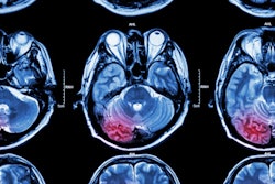
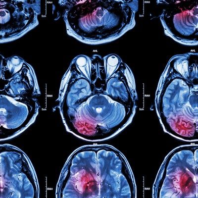
In cases of acute ischemic stroke, imaging of the extracranial arteries is necessary to detect atherosclerosis and potentially associated comorbidities such as carotid artery stenosis and large vessel occlusion or dissections. CE-MRA represents the standard of care in stroke MRI, but it has several risks and limitations, particularly nephrogenic systemic fibrosis, allergic reactions, and the uncertain long-time effects of gadolinium deposition in the brain, the authors explained. Additionally, image quality of CE-MRA may be hampered due to mistiming of acquisition regarding the first pass of the contrast bolus, resulting in venous contamination or insufficient contrast.
Many non-CE-MRA techniques have been developed, with time-of-flight (TOF)-MRA being widely regarded as insufficient for imaging of the extracranial arteries.
Against this background, the researchers decided to evaluate the image quality of 3D REACT (being adapted to this distinct vascular territory) in comparison with standard high-resolution 3D CE-MRA. Their retrospective, single-center study included 35 patients (mean age 60.31 ± 21 years, 17 females) who received a standard MRI stroke protocol (Philips Ingenia 3T, Philips Healthcare) between May and July 2019 in clinical routine.
The protocol included both flow-independent 3D isotropic REACT (compressed SENSE factor 4 for acceleration of image acquisition; fixed scan time, 2 minutes 46 seconds) and high-resolution 3D CE-MRA (compressed SENSE factor 6) of the extracranial arteries.
Three blinded radiologists independently scored 630 arterial segments -- aortic arch/adjacent branches, bilateral common carotid artery (CCA), internal carotid artery (ICA) (C1- and C2-segment), bilateral proximal external carotid artery (ECA), bilateral distal ECA (parotid space), bilateral vertebral artery (V1-, V2, and V3-segment) -- based on three criteria: vessel delineation, vessel signal, and vessel contrast to surrounding tissue.
Overall image noise and artifacts (blurring artifacts, banding artifacts, pulsation artifacts, and parallel imaging reconstruction artifacts) were assessed. For the subjective evaluation of image quality, a five-point Likert scale (1 = nondiagnostic, 5 = excellent/none) was used. For objective evaluation of image quality, an independent fourth radiologist measured bilateral apparent signal- and contrast-to-noise ratios (aSNR/aCNR) for CCA, ICA (C1-segment), and vertebral artery (V2-segment) in comparison to perivascular tissue.
In comparison to REACT, CE-MRA provided better delineation for the CCA and ICA (C1- and C2-segment, each median 5 [min 2 - max 5] vs. 4 [3-5]; P<0.05). For the ICA, REACT achieved higher signal (C1-segment, median 5 [3-5] vs. 4 [2-5], P<0.05; C2-segment: median 4.5 [3-5] vs. 4 [2-5], P>0.05) as well as contrast (C1- and C2- segment: median 5 [3-5] vs. 4 [2-5] (P>0.05)) than CE-MRA.
"The remaining segments of the blood supplying vessels of the brain showed equal medians. For these arteries, no segment achieved a lower minimum score in REACT compared to CE-MRA," the authors wrote.
Key findings and the future
There was no statistical difference for artifacts, whereas REACT yielded a significantly lower image noise (median 4 [3-5] vs. 4 [2-5], P<0.0001) with significantly higher aCNR (51.77 vs. 40.3; P=0.002) and aSNR (58.81 vs. 43.74; P<0.0001) for all vessels combined. For compressed SENSE reconstruction and acquired mDIXON XD images with their different readouts, they noted an average time of 3 minutes and 39 seconds (±25 seconds, ranging from 3 minutes and 8 seconds to 4 minutes and 33 seconds] from start of the acquisition to complete image reconstruction.
Looking to the future, the group's next project is to evaluate the technique's performance in assessing carotid artery disease in a larger cohort of patients.
Editor's note: to view the authors' full e-poster and clinical images, go to the ESR's EPOS website.
Disclosures: One of the study authors, Kilian Weiss, PhD, is an employee at Philips Healthcare. Another author, Dr. Jan Borggrefe, is a speaker at Philips Healthcare.