Diffusion-weighted imaging (DWI) of the breast offers enormous future potential as an imaging biomarker, but multicenter trials are essential to validate single-center studies and establish DWI as a useful clinical decision-making tool, according to Dr. Julia Camps-Herrero, president of the European Society of Breast Imaging (EUSOBI).
In a free podcast produced by the British Institute of Radiology (BIR), she elaborates on the development and validation of diffusion MRI as an imaging biomarker and the potential applications of diffusion-weighted MRI, and also shares her thoughts on the future for breast imaging.
"Integrating imaging biomarkers into clinical practice allows us to measure the underlying pathophysiological mechanisms, noninvasively, revealing tissue properties relevant to detection, diagnosis, prognosis, or response to therapy," noted Camps-Herrero, a breast radiologist and corporate chief of breast health at Ribera Salud, a network of hospitals in Madrid and southeast Spain.
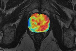
 Breast images obtained with DWI in a patient with a triple-negative subtype cancer in the left breast. (a) Corresponding slices from dynamic contrast-enhanced (DCE) postcontrast image, (b) DWI at b = 0 s/mm2, (c) DWI at b = 700 s/mm2, (d) apparent diffusion coefficient (ADC) map. Invasive tumors show reduced diffusivity on DWI, appearing hyperintense on b = 700 s/mm2 image (c) and hypointense on the ADC map (arrows) (d). All images courtesy of Dr. Julia Camps-Herrero and BJR | Open.
DWI provides complementary information to dynamic contrast-enhanced (DCE) MRI exams. It has already proved its value in single-center studies on lesion characterization and response evaluation but has a less clear-cut role in tumor profiling through prognostic associations. Technical issues due to standard echo-planar imaging (EPI) are gradually being solved, but the technique must be completely standardized, and clear interpretation guidelines must be issued before DWI can become fully incorporated in breast cancer diagnosis and response evaluation, she explained.
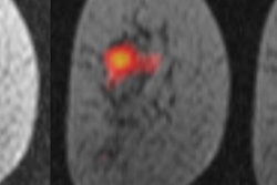
 Same patient as in previous figure. Corresponding slices from (a) DCE postcontrast image, (b) DCE subtraction image, (c) ADC map. Two BI-RADS 4 lesions (arrows) were identified in a different quadrant from the index tumor. ADC characterized them as malignant (ADC = 1.28 x 10⁻³ mm2/sec). An MR-guided vacuum-assisted biopsy was performed in the largest of the two lesions, and an additional multicentric invasive ductal cancer was confirmed.
Future growth potential
Potential areas of future growth include cancer detection using DWI without contrast agents, investigation of additional biological properties through DWI modeling, and analysis of radiomics classifiers in order to better stratify patients and enable a real precision medicine, Camps-Herrero noted.
The high intrinsic contrast achieved with DWI without the injection of external agents, and the ease of implementation into multiparametric protocols due to the fact it is an inexpensive and fast sequence, has triggered several studies on the role of DWI as an alternative to DCE for breast cancer screening. This research has been undertaken in different populations, mostly in patients with suspicious mammographic and ultrasound findings. Because of the issues raised by breast density legislation in U.S., as well as concerns about the long-term use of gadolinium contrast agents, this is a very active area of research with promising preliminary data, she pointed out.
In DWI modeling, the monoexponential decay model is the most commonly used in clinical applications, but to extract additional biological information, more sophisticated techniques are being explored. These techniques require increased acquisition times and complex postprocessing tools, added Camps-Herrero, who is also vice president of the Spanish Breast Imaging Society (SEDIM).
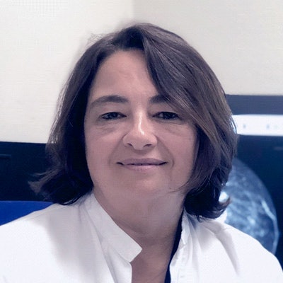
 Dr. Julia Camps-Herrero from Alzira, Spain.
In radiomics, the main objective is the discovery of "signatures" (imaging biomarkers) that have strong associations with a patient's prognosis or tumor subtype classification, she continued. Imaging biomarkers have the potential to capture the entire tumoral area, including its heterogeneity, in a noninvasive and quick way; and in radiomics, a great number of image features -- including shape, histogram, and texture features -- are extracted from medical images.
"One of the most interesting areas of research today is the evaluation of residual disease after neoadjuvant treatment," she said. "Residual disease has been traditionally evaluated with contrast-enhanced MRI, but research shows that diffusion adds sensitivity for predicting a pathologic complete response, which is the holy grail in response evaluation."
Overall, there is still some way to go, despite such promise, Camps-Herrero emphasized, and it is very important for breast radiologists to become better acquainted with this process of implementation, validation, and standardization of imaging biomarkers.
"We have to become very familiar with these new terms," she said. "Diffusion can increase the accuracy of contrast-enhanced MRI in everyday clinical practice. I use diffusion a lot when I am staging a breast cancer and I see additional lesions that I would like to characterize."
Diffusion, abbreviated, and ultrafast MRI techniques, plus other novel developments such as metabolic imaging, hyperpolarized MRI, Mammi-PET, and artificial intelligence, will be addressed at the upcoming EUSOBI annual meeting, to be held in Budapest from 3 to 5 October.
Editor's note: You can read the author's original BJR | Open article on which this podcast was based ("Diffusion-weighted imaging of the breast: Current status as an imaging biomarker and future role"), on the BIR website.