In suspected cases of pulmonary tuberculosis (TB) where radiation dose is of great concern, particularly in younger patients, lung ultrasound is a valid, noninvasive, and cost-effective tool compared with chest CT, new research from Italy has found.
"Most of the alterations found with lung ultrasound were nonspecific and did not allow a differential diagnosis with other pathologic pulmonary conditions. But, once TB alterations had been detected with a chest CT, lung ultrasound could play a consistent role in the follow-up of peripheral lesions during antibiotic therapy," noted lead author Dr. Diletta Cozzi and colleagues from the department of emergency radiology at Careggi University Hospital in Florence, Italy.
Global incidence
TB is a worldwide infection that now kills more than 1 million people a year, they explained in an e-poster due to be presented at the upcoming congress organized by the European Society of Thoracic Imaging (ESTI) and the Fleischner Society. The meeting begins in Paris on 9 May, but the e-posters are available in advance on the Electronic Presentation Online System (EPOS) section of the European Society of Radiology website.
Diagnosis is made through clinical lab findings, but imaging plays an increasing role in the management of these patients. Usually the main radiological approach in these cases is through radiography and chest CT, but Cozzi and colleagues have obtained preliminary results for a cohort of patients with suspected TB infection studied by lung ultrasound. To evaluate the potential of lung ultrasound in the diagnosis and follow-up of TB, they have compared these findings with those from chest CT.
The Florence group's study involved 33 patients (20 men, 13 women) with a median age of 40.5 years. Between October 2017 and May 2018, all patients were admitted to the hospital's infectious disease department with suspected TB, after being evaluated in the emergency department with a clinical lab and radiological evaluation (chest x-ray and chest CT).
"Lung ultrasound investigation was performed by a radiologist within three days after admission," the authors wrote. "Immediately after, a short report on the observed findings, their location, and spatial extension was written. Lung ultrasound images and video clips were then saved on portable memory in order to easily compare them with the CT ones and with the bronchoalveolar lavage (BAL) results."
Main findings
In the study, chest CT was the gold standard. All patients underwent BAL, which confirmed TB infection in 23 out of 33 cases (70%). Lung ultrasound was negative in three out of 33 patients (10.1%) because of the absence of subpleural alterations detectable with an ultrasound exam.
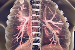
Lung ultrasound was positive in 30 of the 33 patients (90.9%). Among these 30 patients, the researchers found parenchymal-subpleural consolidations in 25 (83.3%), interstitial syndrome (b-lines, comet-tail artifact) in 20 (66.7%), pleural effusion with parenchymal atelectasis in five (16.7%), cavitated areas in six (20%), pleural empyema in one (3.33%), and miliary TB in one (3.33%).
"Only in one patient, we found multiple subpleural micronodules that referred to miliary distribution of TB infection," the authors noted. "The extension of parenchymal involvement detected with lung ultrasound was consistent with the evidence of subpleural alterations in CT in 93% of cases. Upper and middle lobes were often involved. Lower lobes were usually involved in the case of more aggressive disease."
Using only lung ultrasound, it was not possible to distinguish patients with active TB (BAL+) or not (BAL-), they concluded.
Vigilance of radiologists
In another ESTI 2019 e-poster, Korean authors outlined how TB presents a wide spectrum of clinical and imaging findings and may affect many different organs in different ways.
"The diagnosis requires a high degree of [suspicion, and] it is important that the radiologists recognize the imaging findings allowing for the establishment of a more effective strategy to confirm the diagnosis and to institute the appropriate treatment as soon as possible," noted corresponding author Dr. Ki Yeol Lee, a radiologist at Korea University Ansan Hospital, and colleagues.
TB is a public health problem worldwide, particularly among immunocompromised patients and other high-risk groups. It manifests in active and latent forms, and familiarity with the imaging, clinical, and laboratory features of TB is important for diagnosis and management; if left untreated, the consequences can be devastating for the patient, they added.