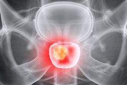
A team of researchers from the U.S. has developed an artificial intelligence (AI) algorithm that could enable less-experienced radiologists to better detect prostate cancer on multiparametric MRI (mpMRI). The group reported the findings at the recent IEEE International Symposium on Biomedical Imaging in Venice, Italy.
The internally developed deep-learning algorithm, called FocalNet, achieved comparable sensitivity to genitourinary radiologists with at least 10 years of experience for detecting prostate cancer lesions on mpMRI. FocalNet also yielded promising accuracy for predicting the lesion's Gleason score (GS).
"As prostate MRI reading quality largely varies according to readers' experience, FocalNet can potentially assist less experienced readers or augment the [prostate cancer] detection task for nonexperts," wrote the team led by first author Ruiming Cao from the University of California, Los Angeles (UCLA).
The research was a runner-up for best paper at the IEEE meeting and was also published online on 27 February in IEEE Transactions on Medical Imaging.
Although mpMRI is often considered the best noninvasive imaging modality for diagnosing prostate cancer, it's currently limited by a reliance on qualitative or semiquantitative interpretation criteria. This leads to interreader variability and a suboptimal ability to assess the aggressiveness of lesions, according to the researchers.
To improve upon this situation, the UCLA researchers sought to train a multiclass 2D convolutional neural network (CNN) to detect prostate cancer lesions and predict their aggressiveness using the GS.
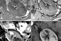
To train the model, the researchers gathered a prostate mpMRI dataset from 417 patients who underwent 3-tesla mpMRI exams prior to receiving robotic-assisted laparoscopic prostatectomy. These mpMRI exams had been prospectively interpreted by three genitourinary radiologists with more than 10 years of experience in reading clinical prostate MRI studies. Radiologist findings of a Prostate Imaging Reporting and Data System (PI-RADS) v2 score of 3 or more were considered to be MRI-positive, while other results were deemed to be MRI-negative.
The 417 patients in the study had a total of 728 annotated lesions, including 286 lesions with GS of 3+3, 270 with GS of 3+4, 110 with GS of 4+3, 30 with GS of 8, and 32 with GS ≥ 9.
After lesion findings were confirmed with whole-mount histopathology, all lesions that were retrospectively judged to be visible on MRI exams were then annotated on each MRI exam. Next, the researchers trained and validated FocalNet on this dataset using fivefold cross-validation.
| Performance of FocalNet algorithm in detecting prostate cancer | ||
| Interpreting radiologist using PI-RADS v2 scoring system | FocalNet (at one false positive per patient) | |
| Sensitivity for index lesions | 93.1% | 89.7% |
| Sensitivity for clinically significant lesions | 89.4% | 87.9% |
The differences in sensitivity between the interpreting radiologist and FocalNet weren't statistically significant. In addition, FocalNet produced an area under the curve (AUC) of 0.81 for classifying clinically significant prostate cancer (lesions with GS ≥ 3+4) and 0.79 for classifying prostate cancer with GS ≥ 4+3.
The research suggests that FocalNet could save time and potentially provide diagnostic guidance to less-experienced radiologists, according to the researchers.