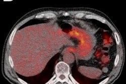
Combining metabolic and morphologic data from FDG-PET/CT scans can significantly improve the detection of lymph node metastasis in patients with muscle-invasive bladder cancer before their surgery, according to a French study published online on 21 January in European Radiology.
The researchers found that the winning formula is to combine CT data on the size of the lymph node and maximum standardized uptake values (SUVmax) from FDG-PET to optimize lymph node staging.
"Combining SUVmax and axial-based lymph node size criteria using FDG-PET/CT improved the diagnostic accuracy for preoperative lymph node staging in patients with invasive bladder cancer, in per area analysis," wrote the researchers, led by Dr. Antoine Girard from the nuclear medicine department at Université Paris-Saclay (Eur Rad, 21 January 2019). "Regional and distant staging is of major importance in the management of muscle-invasive bladder cancer, since accurate staging dramatically impacts treatment efficacy."
FDG-PET/CT's efficacy
The use of FDG-PET/CT to assess lymph node staging in patients with muscle-invasive bladder cancer preoperatively is somewhat controversial, according to the study authors. A few studies have offered evidence that the hybrid modality can detect more distant metastases, compared with CT alone, so there might be the potential for some benefit in managing patients with this disease. Girard and colleagues, however, contend that the "heterogeneous design of these studies did not allow reliable conclusions to be drawn."
Other research has considered various threshold values based on size with CT, such as lymph nodes larger than 10 mm, as well as SUVmax with PET and CT. None of these studies, though, combined both PET and CT data to form any conclusions.
So, the French researchers decided to take that next step and see if SUVmax with lymph node size can improve the detection of regional and individual lymph node metastases and to "compare the accuracy of prospectively acquired FDG-PET/CT to the accuracy of CT alone for preoperative regional lymph node staging in patients with muscle-invasive bladder cancer," they wrote.
Between May 2015 and May 2017, a total of 61 patients (median age, 73 years) were prospectively referred for baseline staging. All patients had no history of pelvic or genitourinary cancer and no distant metastasis on conventional imaging. FDG-PET/CT scans (Discovery 610 Elite, GE Healthcare) were performed prior to neoadjuvant chemotherapy, with the median time of 65 days (range, 2-140 days) between imaging and radical cystectomy.
The scans discovered a total of 1,012 lymph nodes in 122 pelvic areas of the 61 patients. In all, 62 lymph node metastases from bladder cancer were discovered in 17 patients across 24 regions.
How did PET/CT perform?
Relative to staging, lymph node size and SUVmax were significant indicators of malignancy. The median size of malignant lymph nodes was 7 mm (range, 5-9 mm), compared with a median size of 5 mm (range, 5-6 mm) for benign lymph nodes (p = 0.015). Malignant lymph nodes also had significantly higher SUVmax values with a median of 1.9 (range, 1.4-4.3), compared with a median SUVmax of 1.3 (range, 1.0-1.7) for lymph nodes with no malignancy (p = 0.002). In 35 regions, lymph nodes were too small to be detected by CT and PET.
Based on the regional analysis, FDG-PET/CT achieved greater diagnostic accuracy of 84%, compared with CT alone (78%), which reached statistically significant superiority (p = 0.039). In the patient-based analysis, FDG-PET/CT correctly classified pelvic lymph node status in five more patients (8%) than CT alone, for diagnostic accuracies of 82% and 74%, respectively. The statistical difference, however, was not statistically significant (p = 0.13).
| Accuracy of PET/CT compared with PET/CT in lymph node staging | ||
| Regional analysis* | CT alone | PET/CT |
| Sensitivity | 25% | 29% |
| Specificity | 91% | 97% |
| PPV | 40% | 70% |
| NPV | 83% | 85% |
| Accuracy | 78% | 84% |
| Patient-based analysis** | CT alone | PET/CT |
| Sensitivity | 41% | 47% |
| Specificity | 86% | 95% |
| PPV | 54% | 80% |
| NPV | 79% | 82% |
| Accuracy | 74% | 82% |
*Statistically significant difference (p = 0.039) between PET/CT versus CT alone.
**No statistically significant difference.
Nine patients had no visualized lymph node involvement detected by CT or PET. However, pelvic lymph node involvement was discovered in three of those patients on a final pathological exam.
So, how can the results benefit patients with muscle-invasive bladder cancer? After determining which patients have malignant lymph nodes, clinicians can take appropriate therapeutic measures to manage the disease.
"Thus, every effort must be made at time of diagnosis to identify lymph node involvement and provide full-course induction systemic chemotherapy to lymph node-positive nonmetastatic patients," the authors wrote. "The high specificity (≥ 95%) of combined PET/CT to detect malignant pelvic lymph nodes, both on regional-based and patient-based analyses, helps to select the right patients for systemic chemotherapy rather than useless radical cystectomy."
In addition, as more clinicians utilize immunotherapy, FDG-PET/CT should also be "assessed for predicting and monitoring treatment response," they concluded.