Photodynamic therapy (PDT) uses a light-activated drug to create reactive oxygen species that kill nearby cancer cells. Its use is limited, however, by the low penetration of light into tissue and insufficient accumulation of the photosensitizing drug in tumors. Nanoparticles engineered to deliver the photosensitizer can improve its targeting to tumor sites, but drug accumulation is highly variable between tumors and patients.
A team of scientists in Russia has devised a technique that uses MRI to identify the time of peak photosensitizer accumulation in the tumor, enabling irradiation when the concentration of the drug is at a maximum. The team has now proved the effectiveness of this approach in preclinical tests.
The researchers -- from the National University of Science and Technology (NUST MISIS), the Moscow Technological University (MIREA), and the Pirogov Russian National Research Medical University -- created magnetic nanoparticles (MNPs) loaded with photosensitizer molecules. They evaluated the potential of these hybrid particles for PDT of mice bearing colon carcinoma tumors.
After injecting the mice with the photosensitizer-loaded MNPs, the researchers used MRI to track the particles in the animals' bloodstream in real-time and monitor their accumulation in tumor tissue. MRI revealed peak MNP accumulation in the tumors 60 minutes after injection.
The team verified this finding using atomic emission spectroscopy to measure iron concentration and fluorescence imaging to measure drug accumulation in tumor tissue. Importantly, the photosensitizer delivery profile in tumors was consistent with the MNP accumulation dynamics, suggesting that the two components were delivered to the tumor site together and behaved as a single complex in vivo (Pharmaceutics, 17 December 2018, Vol. 10:5, p. 284).
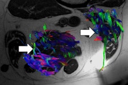
 Maksim Abakumov and colleagues hope to improve the effectiveness of photodynamic therapy. Image courtesy of NUST MISIS.
The researchers treated the animals with PDT at different time points after injection and evaluated the resulting tumor growth curves. "We conducted a series of preclinical tests on three groups of mice for 21 days," explains co-author Maksim Abakumov, head of the NUST MISIS Biomedical Nanomaterials Laboratory. "The first group received radiation 30 minutes after injection of the test drug, the second one at 60 minutes, the third after three hours or more."
Consistent with the MRI-predicted drug accumulation peak, PDT performed 60 minutes after injection was more efficient in inhibiting tumor growth than treatment scheduled 30 or 240 minutes postinjection.
In the first week after treatment, all animals that received PDT demonstrated delayed tumor growth compared with a control group. At day 7, no tumors were found in mice irradiated 30 or 60 minutes after injection, while tumors in the 240-minute group were smaller than those in the control group. From day 10 onward, however, tumor regrowth was observed in the 240-minute group. Animals in the 30-minute group exhibited relapse at day 14, but all animals in the 60-minute group were tumor-free up to day 22.
"Almost all mice from the second group demonstrated a stop in tumor growth, which proved the correctness of the proposed hypothesis," Abakumov said.
The researchers concluded that tracking MNP accumulation using MRI can predict peak drug concentration in tumors, enabling scheduling of PDT to maximize antitumor response. In the near future, the team plans to start clinical trials of the hybrid nanoparticle.
Tami Freeman is the medical and biophysics editor for Physics World.
© IOP Publishing Limited. Republished with permission from Physics World, a website that helps scientists working in academic and industrial research stay up to date with the latest breakthroughs in physics and interdisciplinary science.