This month, June 2018, marks the 60th anniversary of U.K. researcher Dr. Ian Donald's classic article on ultrasound, published in the Lancet. Today, we cannot imagine a hospital without access to ultrasound. Ultrasound has changed medical management and diagnosis for the better, enabling more accurate noninvasive diagnosis without radiation in many medical scenarios, and not just for the obstetric patient.
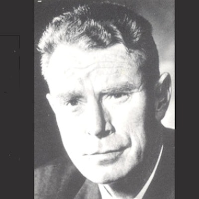
 Dr. Ian Donald was always concerned with the ethical and moral issues in gynecology and obstetrics. All photos courtesy of Dr. Adrian Thomas.
Few people can really claim to have revolutionized medical practice, but Donald is one of them. His legacy is all around us in every country, and has helped make childbirth safer worldwide by enabling accurate antenatal diagnosis so that appropriate care can be tailored for the pregnant mother and the newborn child.
Donald was born in Cornwall, England, in 1910. His family moved to South Africa, where he studied in Cape Town, but in 1930 he returned to London to study medicine at St Thomas's Hospital, where he qualified in 1935. He started his career as a house physician in the obstetrics and gynecology department there. He served as a medical officer in World War II and was awarded a military member of the Most Excellent Order of the British Empire in 1946.
He became a reader in obstetrics and gynecology at the Hammersmith Hospital in London in 1951, a venue that accounted for many of the future leaders in U.K. academic medicine. In 1954, he was appointed to the Regius Chair of Midwifery in Glasgow, Scotland. It was here that he did his groundbreaking work.
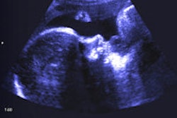
Along with Tom Brown, the engineer form Kelvin and Hughes, he started his experiments with ultrasound, resulting in building the first machine with Brown in 1958. This led to the ultrasound images first published in the 1958 Lancet article written with Dr. John McIver (registrar in the unit) and Brown.
 The one ton (1,016 kg) diasonograph machine was sometimes known as the "dinosaurograph." The heavy gantry was important because of the need for a stable scanning platform. This photo was taken in 1972.
Donald was regarded as an eccentric and a maverick, but continued with his studies on this new imaging technique. The first scanner, the Diasonograph, was built by Fleming and Brown for Kelvin and Hughes and was sold to Sweden in 1961. Early scanners were large and cumbersome and had poor image quality compared with modern standards. Nevertheless, many of these early machines were manufactured and sold in the 1970s by Nuclear Enterprises. Although many were skeptical in the early days, they were soon won over when the clinical uses became more apparent.
 The grave of Ian Donald in Paglesham: "In loving memory, Ian Donald, 1910-1987, Regius Professor of Midwifery, Glasgow, pioneer of medical ultrasound."
Donald was showered with awards, including a commander of the Most Excellent Order of the British Empire in 1973, an honorary doctor of science degree in 1981 from London, the same degree from Glasgow in 1983, an honorary fellow of the Royal College of Obstetricians and Gynaecologists in 1982, and was made an honorary fellow of the British Medical Ultrasound Society in 1984 along with Tom Brown. He died on 19 June 1987.
Today there are millions of patients worldwide who have benefited from his legacy. At the U.K. Radiological Congress (UKRC) meeting at the start of July, there will be a small exhibition loaned by the British Medical Ultrasound Society archive at the British Society for the History of Radiology stand in the exhibition hall accompanying a talk about his life on the opening Monday of the meeting.
Dr. Arpan K. Banerjee is past chairman of the British Society for the History of Radiology and co-author, with Dr. Adrian Thomas, of The History of Radiology.
References
- Donald I, McVicar, J Brown TG. Investigation of abdominal masses by pulsed ultrasound. Lancet, 1958;1(7032):1188-1195.
- Thomas AMK, Banerjee AK. The History of Radiology. OUP; 2013.
- Thomas AMK, Banerjee AK, and Busch U. Classic Papers in Modern Diagnostic Radiology. Springer; 2005.
The comments and observations expressed herein do not necessarily reflect the opinions of AuntMinnieEurope.com, nor should they be construed as an endorsement or admonishment of any particular vendor, analyst, industry consultant, or consulting group.