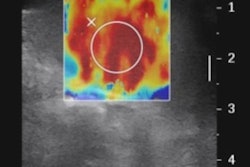
Lung ultrasound finds more cases of pneumonia in children and young adults than chest radiography does, according to a study published online on May 11 in the journal Ultrasound in Medicine and Biology.
Other research has found that lung ultrasound has an overall sensitivity of 96% and a specificity of 93% for identifying pneumonia, and its use for this particular application has been steadily increasing, wrote a team led by Dr. Giulio Iorio of San Giovanni di Dio Hospital in Naples, Italy.
"The use of lung ultrasound for the diagnosis of community-acquired pneumonia has been growing in multiple settings, and numerous studies have reported its reliability and accuracy in diagnosing this pathology in children and young adults," Iorio and colleagues wrote. "[Some] studies [even] promote lung ultrasound as the first choice or as a valid alternative tool to replace chest radiography."
Valid alternative
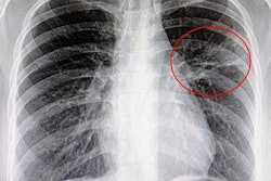
Pneumonia is one of the leading causes of death among children around the world, with 95% of cases occurring in developing countries, according to the researchers. It is usually diagnosed with a patient's clinical history and by tracking the patient's respiratory rate, fever, respiratory signs, and symptoms; x-ray is supposed to be used in severe or complicated cases (Ultrasound Med Biol, May 11, 2018).
But x-ray is regularly performed in children with mild or uncomplicated pneumonia, exposing them to unnecessary radiation, the group noted. Ultrasound offers an accurate and less damaging alternative.
"One of the limitations of radiography is the risk of damage from ionizing radiation, which is greater for children than for adults," Iorio and colleagues wrote. "Numerous studies have reported the high diagnostic accuracy of ultrasonography imaging and indicated that lung ultrasound is an excellent tool for detecting pneumonia in children. For these reasons, lung ultrasound should be the main alternative to x-ray or, at least, the first-line examination."
For their study, the researchers investigated whether there were any differences in the number, position, and size of lung lesions between ultrasound images and corresponding x-ray images in pneumonia cases. They then analyzed the reasons for any differences and whether the differences had clinical implications.
The study included data from 47 children with a final diagnosis of pneumonia between January 2014 and December 2016. The children ranged in age from 1 month to 12 years. The average hospital stay was five days, and the most common symptoms were fever and cough. All of the patients underwent both x-ray and lung ultrasound; the exams were conducted within 24 hours of each other. Both x-ray and ultrasound images were read by a radiologist and a sonographer.
In the 47 cases of pneumonia, chest radiography identified 44 lesions, while lung ultrasound found 72 -- or 28 more, the group noted. Of these 28 lesions, there were 15 consolidations 1 cm or smaller, 12 larger than 1 cm, and one minimal pleural effusion.
The researchers also found the following:
- Three pleural effusions identified on x-ray actually turned out to be lung consolidations on ultrasound.
- One minimal pleural effusion found on lung ultrasound was missed by chest radiography.
- Of the 12 consolidations greater than 1 cm, three were detected in the right lung with both imaging modalities, but x-ray missed nine.
- Chest radiography identified consolidations greater than 1 cm bilaterally in one case, while ultrasound found consolidations in both lungs in seven cases.
The study definitely demonstrates that ultrasound finds lung consolidations that chest radiography misses, and smaller ones as well, according to Iorio and colleagues. Lung ultrasound also found more cases of double pneumonia than x-ray did.
"[False]-negative radiologic findings could mislead the physician both on diagnosis and on therapeutic management because of their failure to recognize pneumonia and double pneumonia," Iorio and colleagues noted.
Another solution
Guidelines for diagnosing pneumonia state that there's no need for children to undergo chest radiography unless their condition is severe, but most who present with symptoms do have an x-ray -- in part because clinical examination is considered unreliable, the authors wrote. Ultrasound could offer a good solution to the problem of x-ray overuse.
"[It's] desirable to modify the workup for the diagnosis of nonsevere community-acquired pneumonia in children ... [and] greater use of lung ultrasound would be appropriate," the group concluded. "Based on the principle primum non nocere, lung ultrasound could represent a first-line tool to be followed by chest radiography if the diagnosis remains unclear."