German researchers have found no evidence of increases in T1 signal intensity among patients who received more than 20 injections of macrocyclic gadolinium-based contrast agents (GBCAs) for MRI scans, according to a study published online December 7 in Radiology.
While it is too soon to draw definitive conclusions on the safe use of macrocyclic GBCAs, the findings shed additional light on the potential effect of gadolinium deposits, which may linger in the brain years after injection (Radiology, December 7, 2016).
 Dr. Alexander Radbruch from Heidelberg University.
"There is currently a debate if the implications of gadolinium deposition in the brain should affect daily clinical practice even without an evidence of a demonstrated adverse clinical outcome," wrote a team led by Dr. Alexander Radbruch from Heidelberg University. "Our study contributes to the debate by providing further evidence that the gadolinium deposition risk might be minimized when macrocyclic GBCAs are used."
Gadolinium retention
In a December 2013 study, Kanda et al brought to light the issue of increased signal intensity in the dentate nucleus after multiple injections of gadolinium contrast, and since then more than a dozen other studies have explored the effect of different GBCAs on signal intensity in the dentate nucleus and other regions of the brain. Several papers showed evidence that such hyperintensities in the dentate nucleus are specifically caused by linear GBCAs.
In fact, a 2016 study by Radbruch et al suggested that switching from a linear GBCA to a macrocyclic GBCA reduced the level of residual signal intensity in patients who underwent multiple enhanced MRI scans with both types of contrast agents. The authors did caution, however, that the "decrease was rather small and the evidence provided in the study is limited."
In the current retrospective study, Radbruch and colleagues narrowed their focus to possible evidence of signal intensity on unenhanced T1-weighted MR images in the dentate nucleus of patients who received an average of 23 injections of macrocyclic GBCAs.
A total of 33 patients were studied. All MRI scans were performed exclusively with 3-tesla scanners (Trio, Verio, Prisma, and Skyra, Siemens Healthineers) using the macrocyclic agents gadobutrol (Gadavist, Bayer HealthCare) and gadoterate meglumine (Dotarem, Guerbet). The mean injected dose per MRI scan was 7.74 mmol for gadobutrol and 12.61 mmol for gadoterate meglumine, based on the patient's body weight. There was an average of 12 weeks between every administration.
Signal intensity ratio differences were calculated based on dentate nucleus-to-pons and dentate nucleus-to-middle cerebellar peduncle (MCP) values by subtracting the signal intensity ratio of the first MRI scan from the signal intensity ratio at the last MRI exam. The mean interval between the first and last MRI scans was 273 weeks.
Ratio results
The researchers found no significant T1 signal increase in the dentate nucleus in any of the patients after a mean 23 serial injections of the macrocyclic GBCAs. The findings were reinforced by the dentate nucleus-to-pons ratio of -0.0032 ± 0.0154 (p = 0.248) and the dentate nucleus-to-MCP ratio of -0.0011 ± 0.0093 (p = 0.521).
"We assessed the dentate nucleus-to-pons and the dentate nucleus-to-cerebellum through ratios," Radbruch said in a presentation at RSNA 2016 about the study. "We did not find any signal intensity increase."
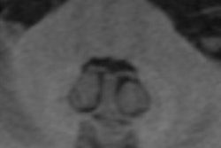
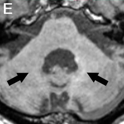
 Axial MR images of a patient before (A) and after nine (B), 17 (C), 29 (D), and 41 (E) administrations of gadobutrol and gadoterate meglumine. Pre-existing hyperintensities in the dentate nucleus (most likely related to prior linear GBCA injection) are visible in all images. There is a slight decrease in hyperintensities between the baseline MRI and the MRI after 41 injections of macrocyclic GBCAs. Images courtesy of Radiology.
As he addressed the issue of gadolinium retention in patients with multiple exposures, Radbruch added that there has been no evidence indicating that any signal intensities have resulted in clinical problems.
"From a methodological point of view, we can come back with as many studies as we want; we can never prove that these hyperintensities, this presumed gadolinium deposition, does not cause some issue," he said. "But it is extremely important to point out that GBCAs can be as lifesaving as medication; no imaging technique can totally replace them. They should be applied only as clinically indicated."