When the doors open to the medical facilities in London's Olympic Village next July, an impressive range of imaging equipment will be on display: two 3-tesla MR systems (or one 1.5-tesla and one 3-tesla), a Discovery HD 750 CT scanner, four Logic E9 ultrasound units, one direct digital radiography machine, and a RIS/PACS with voice recognition. The total value will be around 7 million pounds (7.9 million euros).
"Imaging is felt by the IOC [International Olympic Committee] to be a real premier service for the athletes at games time, so it was the first work stream formed after the successful bid in Singapore," said Dr. Philip O'Connor, imaging lead for the Olympic Games and a consultant musculoskeletal (MSK) radiologist from Leeds, U.K., noting that the other streams are sports medicine, pharmacy services, physiotherapy, emergency services, and the polyclinic. "We are going to deliver a musculoskeletal medical service to the athletes, the Olympic 'family,' and the workforce."
One of the MR systems is likely to have a wide bore to accommodate larger athletes, and negotiations for a dedicated MR unit for the extremities are moving ahead. The CT machine will be used to diagnose stress fractures and for CT-guided interventions. CT was not available at the Beijing Olympics in 2008.
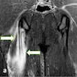
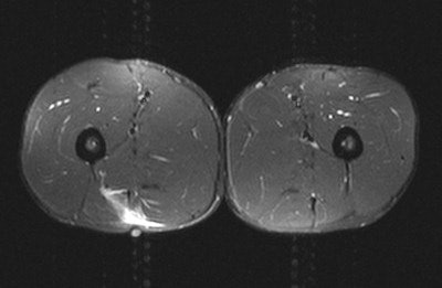 A 28-year-old male sprinter felt sudden onset of posterior thigh pain during a sprint. Swelling, tenderness, and reduced strength were evident. MRI showed grade II myotendinous strain of the long head of biceps muscle, and grade I myotendinous strain of the right rectus femoris muscle associated with scarring of the central tendon. The prognostic indicators were craniocaudal length, percentage cross-sectional area of muscle involvement, presence of an associated scar, and distal versus proximal tears. The clinical diagnosis was a hamstring strain. All images courtesy of Dr. Jeremiah C. Healy.
About 10,000 athletes from 205 countries will participate in the London Olympics, and they will be joined by 11,000 coaches, officials, and IOC members, O'Connor said at a special session held during this month's U.K. Radiological Congress (UKRC) in Manchester. Another 4,000 athletes will take part in the Paralympic Games. In comparison, the last Winter Olympics in Vancouver attracted about 2,500 athletes.
The Olympic Delivery Authority has received approximately 9 billion pounds (10 billion euros) of U.K. government cash to develop the venues that will form the legacy of the games, while the London Organising Committee of the Olympic and Paralympic Games is responsible for organizing the event and is funded entirely by sponsorship. The committee is running the medical services for 62 days in total. Sponsorship packages start at around 10 million pounds (11.16 million euros), and GE is the main sponsor for the imaging center, for which a soft opening is planned on 9 July, 18 days prior to the official opening of the games.
The polyclinic is located centrally within the Olympic Village, but it is next to a railway line and a main road, which is not ideal for setting up MR systems, according to O'Connor. After the Olympics, the building will become a large primary care facility owned by the National Health Service. It's larger than it really needs to be, and medical staff have only had a limited input into the design of the unit, he said. Also, the CT and MR machines will have to be sited in relocatable units outside of the polyclinic.
At London 2012, the workload will be managed by allocating half of the MR slots on a prebooked basis, and having half of them available for walk-in cases. Top priority will be given to athletes who are currently in competition, followed by those in precompetition and postcompetition, coaching staff, officials, Olympic family, and other workers, respectively. The imaging center will be open from 7 a.m. to 11 p.m., and will be staffed by eight radiologists per day (two shifts of four), 16 radiographers (two shifts of eight), and seven radiographic assistants (three for first shift and four for second shift).
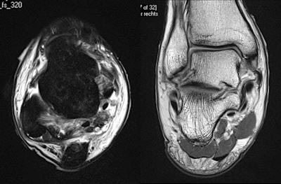 A 24-year professional football player is tackled from the side. The player feels immediate pain in the ankle and is taken off the pitch on a stretcher. Swollen and bruised medial and lateral ligaments and a complex inversion/eversion injury on the fixed foot are apparent. MRI shows grade II lateral and medial ligament injury. Full thickness anterior talofibular ligament tear, partial thickness calcaneofibular ligament tear, full thickness superficial deltoid, and partial thickness deep deltoid can be seen. MRI is the optimal test for integrity of medial and lateral ligaments, but if bony injury is present, CT may be necessary.
Another ultrasound unit will be based at Weymouth, Dorset, where the sailing events will take place. A digital radiography system will be available at Weymouth Hospital, but the nearest MR and CT systems will be in Dorchester. An ultrasound scanner also will be based at Eton Dorney, the venue for the rowing and canoe events. For non-MSK cases, some initial assessments, including chest x-rays, will be done in the accident and emergency section of the polyclinic, but the athletes will be transferred to the nearby Royal London or Homerton University hospitals for further treatment.
At the Beijing Olympics, educational seminars were run on patella tendinopathy and traditional Chinese medicine, as well as a workshop on shockwave examination techniques. A similar program is planned for London.
During the Beijing games, nearly 22,000 medical encounters took place, of which about 45% were workforce-related and only 16% were athlete-related. The organizers performed 450 x-rays, 320 ultrasound exams, and 440 MR exams. The MR service peaked at 59 exams on a single day, which is a heavy workload, O'Connor said.
Of the 858 athlete injuries that occurred in Beijing and for which information is available, nearly 73% occurred during competition, noted Dr. Jeremiah C. Healy, an MSK radiologist at Chelsea and Westminster Hospital in London, at the same UKRC session. About a third of the total injuries resulted from contact with another athlete, 20% were due to noncontact trauma, 20% were due to overuse injuries, 13% were due to contact with an object (e.g., ball or bat), 5.5% were a recurrence of previous injuries, and the remainder were caused by equipment failure or playing/weather conditions. In almost half of the cases, the injury prevented the athlete from competition and training. For more details, he referred delegates to Dr. Astrid Junge's article (American Journal of Sports Medicine, November 2009, Vol. 37:11, pp. 2165-2172).
"There is a great debate about when best to image soft-tissue injuries. The athlete wants it immediately or at least within 24 hours. In my experience, it's better to wait between 24 and 48 hours because otherwise there's a high risk of having a false negative," stated Healy, who helps look after players from several Premiership and Championship football clubs, as well as Premiership rugby union teams, and has been involved in a Champions League thigh-muscle injury study since 2007. "If the imaging does not correlate with the clinical findings, then it's very important to repeat the imaging. Often, if you image too early, then you'll miss the extent of what's going on."
He has a simple rule about which modality to use: If the patient points to the abnormality with one finger, start with an ultrasound, but if the patient points to it with a hand, go straight to MRI. Also, ultrasound is better for imaging superficial structures, while MRI is better for deep structures.
"The general rule is that imaging should be considered only after a provisional clinical diagnosis has been reached and only if it will influence management," said Healy, who performed distant reporting for Team GB, the U.K. Olympic team, during the Beijing games, and is a vice chair of the radiology organizing committee for the 2012 Olympic Games. "However, there is no doubt that coaching staff will influence the need for imaging. ... People get imaged much more often in Northern Europe than they do in Southern Europe, and that may affect what we're expected to provide in terms of service."
Even if the clinical diagnosis is obvious, imaging is still required to gauge the extent of injury and identify any complications. If a significant injury has occurred and there are questions about patient management, then imaging is indicated for preoperative localization. Furthermore, if treatment has failed, imaging is needed to ensure the initial diagnosis was correct, Healy said. Finally, imaging can help document the existence, progression, or resolution of a disease, which can be useful for medicolegal reasons.


