Osteoarthritic cartilage has a different mechanical behavior than healthy cartilage, which may be identified by 3-tesla MRI, according to Austrian researchers. In a new study, osteoarthritic cartilage of the knee tended to display greater deformation upon loading than healthy cartilage, suggesting knee osteoarthritis (OA) affects the mechanical properties of cartilage.
MRI has proved reliable in measuring cartilage thickness and loss in knee osteoarthritis in other studies. However, no previous study has compared the magnitude and pattern of in vivo cartilage deformation between healthy and osteoarthritic knees using quantitative data from MR imaging.
Dr. Sebastian Cotofana, from the Institute of Anatomy and Musculoskeletal Research at the Paracelsus Medical University in Salzburg, Austria, in coordination with a team from the University of California, San Francisco in the U.S. used an MRI-compatible in vivo loading device to study the regional and subregional pattern of cartilage deformation in knees with and without signs of radiographic OA (European Radiology, March 29, 2011).
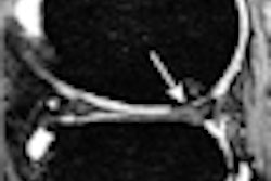
The researchers looked at one knee each in 30 women (11 healthy and 19 with medial, but not lateral, compartmental radiographic femorotibial OA) using 3-tesla MRI. They used a coronal fat-suppressed spoiled gradient-echo (SPGR) sequence. Regional and subregional femorotibial cartilage thickness was determined under unloaded and loaded conditions with 50% body weight applied to the knee in 20° knee flexion during imaging.
Cartilage became significantly thinner during loading in the medial femorotibial compartment (medial tibia -2.7%, medial weight-bearing femur -4.1%), but not in the lateral compartment, where no presence of OA was previously detected, according to the researchers.
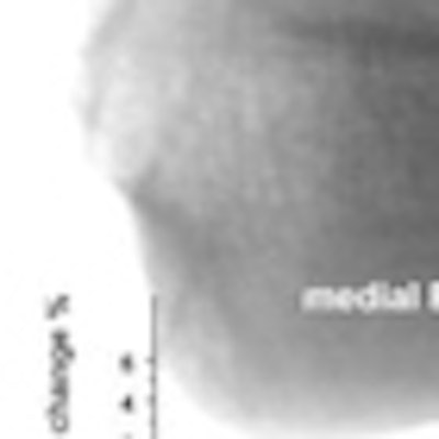
 This image shows the 16 femorotibial subregions with the change in percent (before/after loading) in mean cartilage thickness upon loading with the MRI-loading device applying 50% of the participant's body weight to the investigated knee. Image courtesy of Dr. Sebastian Cotofana.
The magnitude of deformation in the medial tibia and femur tended to be greater in osteoarthritic knees than in healthy knees, the researchers wrote. The pattern of in vivo deformation indicated that cartilage loss in osteoarthritis progression is mechanically driven, they noted.
"Once affected by osteoarthritis, cartilage displays and behaves differently from healthy cartilage, which can lead to more negative impact on cartilage structure and to more deterioration of it," Cotofana said in an interview with AuntMinnieEurope.com. "When physicians understand that cartilage is 'ill,' but still looks healthy, they could potentially start earlier with an adequate treatment -- like weight reduction for instance. The radiologist could help in a very substantial way in the detection of osteoarthritis."
Using 3-tesla MRI gives a better contrast/signal-to-noise ratio, and other tissues such as menisci, synovium, and Hoffa's fat pad can been seen accurately, according to Cotofana.
"I think that more investigations need to be performed in the field of in vivo measures under conditions that are close to daily life conditions like loading," he said. "Maybe some new devices will be developed where we can measure the femorotibial joint in 'action' to understand better what drives progression in osteoarthritis."
