SINGAPORE -- PET/MRI is emerging as a viable tool for pinpointing locations of pain in people with chronic low back pain and persistent hip pain after total hip replacements, according to research by a group from Erasmus MC in Rotterdam, the Netherlands.
Marijn Mostert, a doctoral student, presented a study May 9 at the International Society for Magnetic Resonance in Medicine (ISMRM) meeting showing that F-18 FDG-PET/MRI scans revealed previously unidentified pain locations in 27 patients, with the findings leading to improvements in their care.
“Chronic low back pain and persistent hip pain after total hip replacement are often inadequately treated since conventional imaging techniques such as CT or MRI are unable to accurately identify the source of pain,” Mostert said.
Chronic pain is among the top reasons for visits to physicians worldwide, with up to 43% of chronic noncancer pain patients reporting not receiving treatment. Hybrid PET/MR imaging could help turn this tide over CT and MRI alone, given that it reveals inflammation on a molecular level (PET) combined with highly detailed soft tissue contrast.
To explore its utility, the researchers enrolled 27 patients with unknown pain sources despite previous imaging, diagnostic procedures, or treatment: 15 with low back pain (LBP) and 12 with pain after total hip replacements (THR). The patients were referred to the study by their physicians.
The researchers split the participants into two groups, a control arm (n = 14) consisting of standard clinical care without F-18 FDG PET/MRI or an intervention arm (n = 13; 6 LBP patients and 7 THR patients) who underwent the hybrid scans and subsequent clinical management. Potential pain generators were identified by increased F-18 FDG radiotracer uptake on PET combined with anatomical abnormalities on MRI.
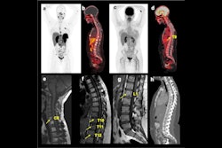
 A 47-year-old female patient with chronic pain in the left lower back/buttock with unknown cause. PET demonstrated focally increased FDG uptake at the left facet L3-4 l. MRI shows minimal fluid, but no edema. Follow-up PET-MRI after six months shows a similar pattern. The patient is on the waiting list for targeted facet joint infiltration (typically not covered by insurance in the Netherlands).Image courtesy of Marijn Mostert
According to the findings, PET/MRI provided new diagnostic insights into the potential pain generator in 12 patients (86%). Moreover, the findings were discussed with the referring physicians, which led to multiple changes in clinical management. Two patients subsequently underwent lumbar nerve root injection, one patient was referred for facet joint injection, and one patient underwent surgical discectomy.
“In our early experience, F-18 FDG PET/MRI can reveal previously unidentified pain generators in patients with chronic LBP or persistent hip pain after THR,” Mostert said.
Ultimately, the group plans further evaluation in a larger cohort to determine the added value of F-18 FDG-PET/MRI in terms of patient outcomes, he added.
“The next crucial step will be to evaluate the impact on patients’ clinical outcomes,” Mostert concluded.