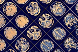
SINGAPORE - Using machine learning with MRI radiomics features accurately assesses brain aging, according to research presented May 6 at the International Society of Magnetic Resonance in Medicine (ISMRM) meeting.
Presenter Eros Montin, MD, of New York University Grossman School of Medicine in New York City reported that a machine learning model using radiomics features from T1- and T2-weighted MR images estimated adult subjects' age with a mean absolute error value of 4.7 years.
The study results could translate to improved clinician understanding of brain changes caused by both healthy aging and those caused by neurodegeneration, Montin noted.
"A machine-learning model capable of accurately estimating brain age could have a large clinical impact," he said.
Previous research on predicting brain age using structural imaging has shown mean absolute error values of between five and seven years, while studies that combined structural and functional imaging information have shown mean absolute error values of less than four years. But functional imaging -- such as functional or diffusion-weighted MRI -- may not be widely available, and prediction accuracy is reached only with large amounts of data (that is, more than 23,000 cases).
That's where MRI radiomics come in. Culling quantitative features from MRI exams "has emerged as a powerful tool for improving patient outcomes and advancing precision medicine," Montin said.
"Radiomics extracts image features from specific regions of interest and then uses machine-learning models to associate features with clinical outcomes," he explained. "For this reason, radiomics requires fewer instances compared with other models trained on the full image, which reduces the degrees of freedom that the model needs to learn."
Montin and colleagues used data from the Human Connectome Project-Aging in a study that included T1- and T2-weighted brain images from 716 healthy adults; this data was used to gather 18,324 radiomics features from areas of the brain understood to be highly affected in the course of normal aging: the bilateral hippocampus, the putamen, and the caudate and to train a stacking regressor machine-learning model (a machine-learning technique that combines predictions of multiple estimators, Montin said). The model included eight regressors: Lasso, random forest, k-nearest neighbors, gradient boosting, AdaBoost, HistGradientBoostingRegressor, and MLPRegressor.

The predictive model using the stacking regressor was trained on only 20 radiomic features --which "supports our hypothesis that these three main brain subcortical areas are enough to provide key information for machine learning-based aging prediction," Montin's team noted -- and estimated brain age with a mean absolute error value of 4.7 years.
"Our study shows that radiomic features can be used for the prediction of brain age in healthy adults with a performance comparable to that reported for models trained on considerably larger datasets available in the literature," Montin said.
The investigators plan to continue this research with muscular sclerosis patients to explore whether the model plus MRI radiomics shows a correlation between brain age and number of brain lesions, according to Montin.