U.K. researchers have reported early success in the development and testing of a high-resolution, compact, hybrid gamma/optical camera designed for intraoperative imaging.
The device is the result of a collaboration between the University of Leicester, the University of Nottingham, and Queen's Medical Centre in Nottingham. The compact gamma camera is the brainchild of researchers at the University of Leicester's Space Research Centre.
"The end goal of this imager is to take it into surgery to be used intraoperatively," said Sarah Bugby, a researcher at the University of Leicester. "We are not quite there yet, but how far we've come so far acts as a good proof of concept."
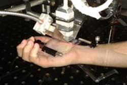
 The high-resolution, compact, hybrid gamma/optical camera in use. Image courtesy of Sarah Bugby.
As the authors noted, the development of portable gamma cameras offers new applications for intraoperative imaging to enhance surgery, and hybrid imaging might assist surgeons in localizing the site of uptake in procedures, such as sentinel node detection.
Technical specs
The compact gamma camera features a 0.5 mm tungsten pinhole collimator and a cesium iodide scintillator-based detector 8 mm x 8 mm in size. The photon detector is an electron-multiplying CCD, which is designed to produce very low noise levels. The handheld device can be used in a portable mode at a patient's bedside or clamped to a stationary object for steady imaging. The unit weighs approximately 1 kg (2.2 lbs).
"We think we've come up with an elegant and quite simple solution for the hybrid camera," Bugby told attendees earlier this month at the annual meeting of the Society of Nuclear Medicine Molecular Imaging (SNMMI). "We are particularly interested in using this in sentinel lymph node biopsies."
Researchers configured the hybrid device by placing a mirror at a 45° angle in front of the pinhole collimator and the optical camera off to the side. The gamma photons from the source pass directly to the mirror, are deflected, and then detected by the optical camera.
"This setup allows us to exactly match the field-of-view of the optical and gamma cameras," she explained. "It doesn't matter what distance you're looking at or what you do with the camera, the images will always line up."
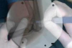
Researchers tested the hybrid system for spatial resolution, energy resolution, and sensitivity based on National Electric Manufacturers Association (NEMA) protocols adapted for use with high-resolution, small field-of-view systems. Image contrast and accuracy of image alignment were evaluated with custom designed phantoms.
Detection accuracy
Bugby and colleagues used custom-made phantoms to simulate a sentinel lymph node biopsy scenario and investigate how detection would be affected by the depth of a node through 100 mm of scattering material and using a node 2 mm in diameter containing 2 MBq of technetium-99 (Tc-99m).
In terms of detection, researchers were able to see abnormalities as small as 1 mm in phantom testing.
At a depth of nearly 13 mm in the phantom, the nodes could be detected from the needle injection site. As researchers moved further away or deeper into the phantom to approximately 6 cm of scattering material, the hybrid device still detected the closest node, which was separated by only 10 mm from the injection site.
At 9 cm, however, scanner material and a loss of sensitivity became "a bit of an issue," Bugby said, and researchers could no longer separate the closest node.
"Obviously, this work is not complete yet," she added. "We are currently looking at far higher activity ratios for what is typical in a sentinel lymph node study and we are also looking at 3D effects."
Image alignment
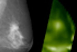
Researchers also evaluated the alignment of the two devices across a phantom by taking the gamma camera image and the optical camera image at the same time to combine them into a hybrid image. The study found excellent alignment between the two technologies, which found the abnormality where researchers expected to see it. "This alignment is really robust," Bugby said.
Under the proposed imaging protocol, clinicians would take the optical image and overlay the gamma camera image. "You can see visually exactly where the activity is compared to what you can see with your eyes," she said. "You can look at that image ... and compare it to the patient in front of you. We think that is what will really aid surgeons in cases like sentinel lymph node biopsies, which can be difficult to exactly direct the radiation."
In the first round of clinical testing on patients, clinicians were able to localize activity of a syringe in the foreground and the site of the injection from a distance of 25 cm in one person undergoing lymphatic imaging, "which was a promising result," Bugby added. "We hope to aid surgeons and clinicians in helping to understand what they are seeing with their gamma camera image."
The researchers are very encouraged with the results so far and plan to continue their study in the clinical setting.
Research funding for the project was provided by the Science and Technology Facilities Council.