Using a stereoradiography (SR) system on the lower limbs in a standing position to calculate patients' morphologic and static parameters can result in a much lower x-ray dose to the ovaries and testes than with CT, French researchers have found.
"In a single acquisition period, this 3D SR system also determined the valgus and varus measurements of the weight-bearing lower limbs. This too should reduce the patients' medical irradiation a little more, by avoiding the need for other medical imaging," noted lead author Dr. Cyrille Delin, from the Réseau d'Imagerie Médicale Maussins-Nollet in Paris. "The use of the SR system to study the torsion of lower limbs makes it possible to reduce the amount of medical irradiation that patients accumulate."
The SR system delivered substantially lower doses of ionizing radiation than CT to all the organs studied. CT doses were 4.1 times higher to the ovaries, 24 times higher for the testicles, and 13 to 30 times higher for the knees and ankles, Delin and colleagues wrote in an article-in-press published online by the European Journal of Radiology on 7 November.
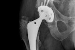
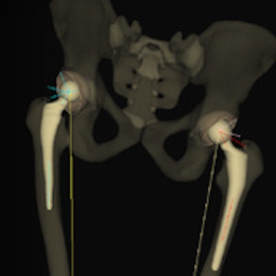
 An example of 3D modeling performed with the EOS system for the assessment of total hip arthroplasty. Image courtesy of Dr. Cyrille Delin.
Relatively few published studies have sought to optimize the dose received by patients during exploration of the lower limbs, particularly the hips, so thestudy team sought to calculate and compare the doses of ionizing radiation delivered to the organs by CT and SR during measurements of lower limb torsion and anteversion.
"Orthopedic specialists and radiologists have long used CT to take these measurements. Its advantages include ease and speed of performance, combined with the wide availability of equipment. CT examination also provides a look at bone morphology and at the quantity and quality of the bone matrix. Its greatest disadvantage, however, is its irradiation, especially of the pelvis and genital organs," the authors explained.
A Rando anthropomorphic phantom (Alderson Rando phantom, Alderson Research Laboratories) was used for the dose measurements. The doses were delivered by a Somatom 16-slice CT scanner (Siemens Healthcare) and an EOS SR unit (EOS Imaging). For the CT scanner, the protocol called for the helical acquisition of the anteroposterior scout (digital) view, focused on three areas of interest: the coxofemoral joints and entire femoral necks, knees (femoral condyles and tibial plateaus), and ankles (malleoli).
Doses to the surface and deeper layers were calculated with thermoluminiscent GR207P dosimeters. Dose uncertainties were evaluated and assessed at 6% at k = 2 (i.e., two standard deviations), according to the authors.
| Absorbed doses for the principal organs assessed | ||
| Organ | SR | CT |
| Ovaries | 0.1 mGy (right), 0.5 mGy (left) | 1.3 mGy (right), 1.1 mGy (left) |
| Testes | 0.3 mGy (right), 0.4 mGy (left) | 8.5 mGy (right), 8.4 mGy (left) |
| Knees | 0.4 mGy (right), 0.8 mGy (left) | 11 mGy (right), 10.4 (left) |
| Ankles | 0.5 mGy (right), 0.8 mGy (left) | 15 mGy (both) |
"Lower limb torsion values (anteversion of femoral necks or torsion of the knees or legs) play a role in the functioning of these limbs and in the development of osteoarthritis of the hip," they stated. "These values must be measured before placement of hip or knee prostheses but also before osteotomies for femoral or tibial correction of bone calluses or dysmorphisms. They are also useful before surgical revision of hip and knee prostheses in cases of poor positioning, given the frequent imprecision of surgeons' intraoperative evaluation of prosthetic femoral anteversion."
The study had important limitations, however. First, the authors could have reduced the irradiation by the SR system still further by using shields for radioprotection of the ovaries and testes, or by placing the phantom with its back to one of the sources, thus increasing absorption of one of the two x-ray beams, required to cross a thicker layer of tissue to reach the genital organs. Nonetheless, the morphology of some patients can make shield positioning difficult.
Similarly, positioning the patient's back to one of the two sources is not always possible, for the morphology of some patients makes it difficult to control their exact placement in the machine. The researchers chose to place the phantom in the SR apparatus in a position applicable to all patients, regardless of morphology.