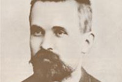
When Wilhelm Conrad Röntgen discovered x-rays in 1895, he observed their effect on photographic glass and fluorescent salts. Certain crystalline salts such as barium platinocyanide or calcium tungstate show fluorescence when exposed to x-rays. These fluorescent effects could be observed by holding a coated screen in front of an object.
It was Thomas Edison who had the idea of placing a tapering box over the screen, with the opening covered by some soft, dark material to fit the face closely and to exclude light. An object placed in front of the screen will then be shown clearly using x-rays. This hooded box, or cryptoscope, was particularly valuable for looking at the chest and thicker parts of the body.
Objects showed up as a black shadow on the fluorescent screen, and this includes the operator's hand when it was pressed against the screen in front of the x-ray tube. By means of a strongly active x-ray tube, the cryptoscope would enable the operator to see the bones in the hands clearly even when the person stood 3 m or more away from the x-ray tube.
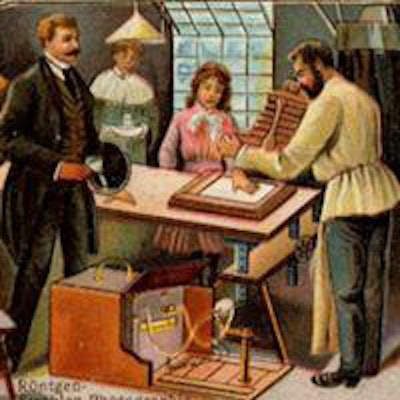
 Fig 1: Use of a cryptoscope depicted on a trade card circa 1900.
The cryptoscope is charmingly depicted on a trade card issued from about 1900 (figure 1). As can be seen in the image from 1896 (figure 2), the danger of the cryptoscope in the early years was related to the lack of protection around the x-ray tube, the lack of protection of the operator, and the habit of the early radiologists to use their own hand as a text object to check the adequate functioning of the x-ray tube.
While Edison did not develop any injuries, his assistant Clarence Dally was not so lucky. Dally developed severe changes in his hands related to the use of the cryptoscope, and sadly he died in 1904 as a radiology martyr after prolonged suffering. Edison stopped working on x-rays because he thought they were too dangerous.

 Left: Fig 2. The use of the cryptoscope (Meadowcroft 1896). Right: Fig 3. The cryptoscope.
Figure 3 shows a cryptoscope in my collection. For obvious reasons, I have never tested it to see that works!
A significant cause of the injuries of the early radiologists was related to the use of the cryptoscope. As has been noted, often the hands were injured, but exposed parts of the body were also affected with x-ray dermatitis. The cryptoscope could be held in the hand, but an operating cryptoscope was made for the use by surgeons in the operating room. The cryptoscope was strapped to the head and could be tilted to allow either inspection of the fracture or fluoroscopy of the fracture to assist in reduction. Despite the dangers, the cryptoscope remained in use for many decades into at least the 1950s. Presumably this was partly related to its ease of use.
Figure 4 shows an advertisement from the British Journal of Radiology in 1942 advertising the Victor handheld cryptoscope. This one had a lead glass window and lead rubber protection for the handler, including the face and hair. The apparatus had a 10 x 8-inch (about 25 x 20-cm) zinc sulphide fluorescent screen and cost a little more than 15 pounds (17.4 euros). This device is advertised as being able to fasten to the head.
','dvPres', 'clsTopBtn', 'true' );" >
It is always interesting is to observe how pioneers rapidly realize the uses to which new technology can be applied. By 1897, the cryptoscope was in use in a Paris railway station to examine luggage for contraband and harmful material (figure 5). Again, note the complete lack of electrical and radiation protection of the x-ray tube and absence of protection to the operator and viewers.
 Fig 5. Examination of a case in a Paris railway station (L'Illustration - 3 July 1897).
Our machines are now much more sophisticated, but the basic principles remain the same. These principles only changed significantly with the introduction of CT scanning in the 1970s.
Dr. Adrian Thomas is chairman of the International Society for the History of Radiology and honorary librarian at the British Institute of Radiology.
Further reading
Thomas AMK, Banerjee AK. The History of Radiology. Oxford, U.K.: Oxford University Press; 2013.
The comments and observations expressed herein do not necessarily reflect the opinions of AuntMinnieEurope.com, nor should they be construed as an endorsement or admonishment of any particular vendor, analyst, industry consultant, or consulting group.