Photon-counting detectors based on cadmium telluride (CdTe) and cadmium zinc telluride offer many advantages for x-ray imaging. Their energy discriminating properties improve material decomposition, eliminate electronic noise, and can reduce patient dose by increasing the image contrast-to-noise ratio, but detectors are hindered by a limited count rate relative to the photon flux used in clinical x-ray and CT systems.
If low-count-rate detectors are used with high-photon-flux x-rays, pulse pile-up effects can occur, where the recorded signals overlap. This leads to distorted energy spectra and unexpected information appearing in the acquired images. Previous studies in this area have focused only on the impact on the energy spectrum. Now, researchers from Yonsei University in Korea have performed a detailed investigation of the effect of photon flux on both the spectrum and the acquired image (Physics in Medicine and Biology, 21 July 2013, Vol. 58:14, pp. 4865-4880).
Reducing the flux
The researchers studied a photon-counting CdTe x-ray detector with 16,384 pixels, used with a microfocus x-ray source. To control the photon flux, they inserted aluminum filters of 1-, 10-, 20-, 30-, and 40-mm thicknesses into the beam.
 Phantom images acquired at 50 kVp with 1- (a, f), 10- (b, g), 20- (c, h), 30- (d, i), and 40-mm (e, j) thicknesses of PFC filters. The upper and lower rows include and exclude electronic noise, respectively.
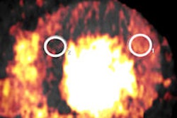
This setup was used to image a cylindrical PMMA phantom containing four holes filled with varying concentrations of iodine-based contrast agent. The researchers acquired energy spectra and images at three x-ray tube voltages (50, 70, and 90 kVp) for the five filter thicknesses. The tube current was set at 10 µA for all acquisitions and the data acquisition time was adapted to acquire roughly the same number of counts for the different filters at each tube voltage.
Image impact
Energy spectra recorded for the different combinations showed a clear dependence on photon flux. With high photon flux, pulse pile-up effects were observed, while for low photon flux, spectra were not affected by pulse pile-up but were slightly impacted by electronic noise. Images created using the distorted spectrum information were also affected.
At 50 kVp, energy spectra measured in the 28-74 keV window revealed that the number of photon counts recorded above the maximum photon energy of 50 keV (i.e. those due to pulse pile-up) did not vary greatly with filter thickness. This implies at this tube voltage, 1 mm of filter was sufficient to reduce the photon flux enough to give negligible pulse pile-up.
 Energy spectra acquired at 90 kVp with five thicknesses of PFC filters.
Images acquired in this energy window showed higher levels of electronic noise with the thicker PFC filters. The researchers note the reduced photon flux does not cause an increase in electronic noise, rather the detectability of the noise increases. Images acquired in the 30-74 keV window were of higher quality, due to the removal of this additional detected noise (which is seen below 30 keV).
The researchers also calculated the signal-difference-to noise ratio (SDNR) between iodine and the background material in each image. Calculated SDNR values for all iodine concentrations were highest with a 10-mm filter, and then decreased gradually as the filter thickened.
','dvPres', 'clsTopBtn', 'true' );" >
At 70 kVp, energy spectra recorded in the 28-98 keV window showed pulse pile-up effects for the 1-mm PFC filter. This reduced as filter thickness increased. Both the image contrast and noise level increased continuously when the photon flux was reduced by thicker filters. The SDNR between the background and the iodine decreased proportionally to the filter thickness, except at 1 mm, with the highest value seen for the 10-mm filter.
Energy spectra acquired at 90 kVp showed the effects of photon flux most clearly, exhibiting a more serious pulse pile-up effect than at 70 kVp. For the thinnest filters, low contrast materials such as PMMA could not be distinguished due to the presence of pulse pile-up.
Spectra with 20- to 30-mm filters eventually showed a reduced number of photons above 90 keV. The images again showed simultaneous increase in contrast and noise when a thicker PFC filter was used, with the highest SDNR measured with a 30-mm filter for most iodine concentrations.
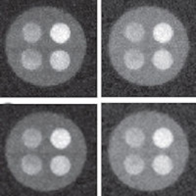
 Phantom images acquired at 90 kVp with 1- (a, f), 10- (b, g), 20- (c, h), 30- (d, i), and 40-mm (e, j) thicknesses of PFC filters. The upper and lower rows include and exclude electronic noise, respectively.
The results of this work demonstrate that photon flux affects both the acquired energy spectra and the resulting images. For all configurations, SDNR was highest at the point where the acquired energy spectrum included the lowest electronic noise without pulse pile-up. In such optimal photon flux conditions, the SDNR was improved by up to 30-fold.
The authors conclude the results demonstrate the possibility of using low-count-rate detectors to perform energy-resolved x-ray imaging with acceptable image quality by using filters to change the photon flux.
© IOP Publishing Limited. Republished with permission from medicalphysicsweb, a community website covering fundamental research and emerging technologies in medical imaging and radiation therapy.