Ghost imaging generates images by analyzing correlations between two light beams, the more powerful of which doesn't bounce off the object in question. The technique has already been demonstrated at visible and infrared wavelengths, but now two groups of scientists -- one in Australia and Europe, and the other in China -- have extended it to the x-ray region. The new results could lead to new methods for medical diagnoses with lower doses of x-rays, or for x-ray crystallography of noncrystalline materials, the researchers said.
Ghost imaging involves splitting a light beam into two beams. The "object beam" is directed toward the object to be imaged, which has a single pixel "bucket" light detector behind. Meanwhile, the "reference beam" travels straight to a multipixel light detector. The idea is to build up a shadow image by incorporating the output from only some of the pixels in the reference detector: those for whom the corresponding parts of the bucket detector are not blocked by the object.
To do this, the detectors are exposed to a broad "speckled" beam whose cross-sectional intensity distribution varies with time. The changing correspondence between the total intensity recorded at the object detector and the pattern of intensities recorded at the reference detector then allows the image to be reconstructed.
Turbulent vision
Ghost imaging at visible wavelengths is already being studied to improve remote imaging of the Earth's surface by satellites in turbulent conditions. Turbulence scatters light in random directions, which usually makes images noisier. But by measuring the intensity correlations of two correlated beams over an extended period of time, one of which is bounced off the object being observed, the effects of turbulence can be averaged out to almost zero.
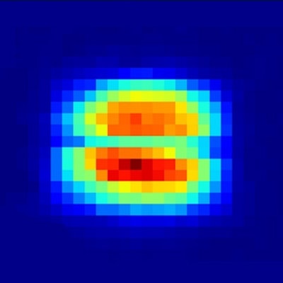
 Ghost image of a wire taken by Daniele Pelliccia and colleagues. The wire runs horizontally across the spot, which is 1 mm across. Courtesy: Daniele Pelliccia et al. Phys. Rev. Lett.
There is another advantage of ghost imaging that also makes it attractive at x-ray wavelengths: A good image can still be generated even when the intensity of the object beam is low. This could potentially significantly reduce the size of x-ray doses administered to patients. However, the difficulties involved in building x-ray optics mean that it is far harder to split x-ray beams than it is beams of visible light.
Daniele Pelliccia, PhD, of RMIT University in Victoria, Australia, formerly at University of Rome "La Sapienza" in Italy, and colleagues got round this problem by using x-rays from a synchrotron source -- ESRF in Grenoble, France. They directed the synchrotron beam at a sliver of silicon, which left part of the beam undisturbed and diffracted the remainder through a small angle. By placing the object -- a copper wire -- in one of the beams and then using different portions of a single camera to serve as both object and reference detectors, the researchers were able to generate ghost images of the wire (Physical Review Letters, 9 September 2016, Vol. 117:11, pp. 113902).
Shot noise
The speckle needed in the experiment was generated naturally by the very short pulses of electrons used to produce the x-rays; the distribution of electrons in each bunch being random and yielding what is known as shot noise. To make sure they really had generated ghost images, the team varied the frequency over which they analyzed the data. They found, as expected, they were only able to produce good images when the frequency matched the electron pulse rate. Failure to image at about one pulse per frame means losing the correlation between the two copies of the beam, Pelliccia explained.
Pelliccia said he and his colleagues are now working out how to exploit this result in order to reduce the dose given in x-ray diagnoses "by at least an order of magnitude" over current levels. Doing so, he points out, will require quite a different technique to the one they used in their experiment, given that synchrotron sources are unsuited to routine medical imaging. In fact, he says, the other new study reporting x-ray ghost imaging, by Shensheng Han of the Shanghai Institute of Optics and Fine Mechanics and colleagues, may point to a more practical alternative.
That research also used x-rays generated by a synchrotron source -- the Shanghai Synchrotron Radiation Facility -- but did not involve beam splitting. Han and coworkers used a single beam directed at a solitary CCD detector, and instead used an actuator to move the object -- in their case a thin gold film with five slits -- into and out of the beam.
To generate the speckle pattern, the researchers placed another gold film, much wider than the beam diameter and containing randomly distributed holes, between the x-ray source and the object. They found they could produce ghost images by moving the holey film across the beam so as to present a continually changing speckle pattern, and then moving the object into and out of the beam once for each different pattern.
Fourier technique
Pelliccia points out the Chinese group didn't generate images directly. Rather, they measured diffraction patterns from the object and then converted those patterns using a Fourier technique. But he says the fact they created their speckle patterns simply by placing a film in the beam, rather than relying on electron shot noise from a synchrotron, makes it potentially better suited to medical imaging than his group's technique. He notes moving a patient in and out of an x-ray beam "wouldn't be a practical option" but adds in future it might be possible to move the detector instead of the patient.
In a paper describing their work, Han and colleagues emphasize the importance of their research to crystallography (Phys. Rev. Lett., Vol. 117, pp. 113901). They say that scientists in future could use their technique to create high-resolution images of noncrystalline samples using "widely accessible laboratory x-ray sources" rather than having to rely on synchrotron sources or free electron lasers. "The structure information of many important molecular materials, such as membrane proteins, is still out of reach because these materials are difficult to grow into macroscopic crystals," they wrote.
Ivan Vartaniants of the DESY laboratory in Germany praises the two groups for their "excellent and challenging work." However, he said the approach put forward by Pelliccia and colleagues suffers from a number of shortcomings, including the current lack of suitably fast and high-resolution detectors as well as the fact that -- unlike copper wire -- biological samples will transmit most of the radiation that passes through them.
As regards the technique developed by the Chinese group, Vartaniants queries whether it could be used to image arbitrarily shaped objects and questions whether it really would be suited to conventional x-ray sources, given that the group tested it using a highly coherent, strong flux synchrotron source.
This article first appeared on physicsworld.com. © IOP Publishing Limited. Republished with permission from physicsworld.com