Portugal is an amazing country with a long history of exploration and innovation going back to the days of Henry the Navigator (1349-1460). In the 1920s, classical radiology was established, and this remarkable era is worthy of close scrutiny.
Many of the more invasive diagnostic techniques developed by the early Portuguese pioneers have been replaced by modern noninvasive methods, but we should salute those who have made enormous contributions to our specialty of diagnostic radiology.
Are historical changes the result of the action of individuals of genius and destiny? Or are there social and cultural trends? And would the discoveries and developments have happened anyway because the time was right? Actually, I think it's not a question of either/or but rather of both/and. We have individuals of genius within a historical stream.
From the earliest days of the use of x-rays, in vitro or postmortem angiograms had been performed. Angiography by either a direct puncture or cut down was achieved with the relatively toxic sodium iodide and bromide solutions in the early 1920s. The major breakthrough in angiography was achieved on 28 June 1927 in Portugal at the Santa Marta Hospital in Lisbon, when the first successful human carotid arteriogram was performed.

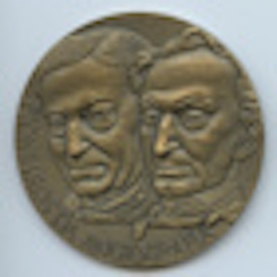
 Fig. 1a and 1b: Egas Moniz and Reynaldo Dos Santos featured on a special commemorative medal in 1982-83.
The two key names are those of Egas Moniz (1874-1955) and Reynaldo dos Santos (1880-1970), as shown on the medal struck in 1982 for a special exhibition (fig. 1a and 1b). In the same year, José Veiga-Pires and Ronald Grainger reviewed their outstanding contribution and that of his Portuguese colleagues in the development of angiography.
 Fig. 2: Egas Moniz appeared on a Portuguese stamp in 1974.
Moniz, a professor of neurology, was the charismatic leader of this Portuguese team and he was celebrated on a Portuguese stamp in 1974 (fig. 2). He was a brilliant polymath, being an author, researcher and clinician, politician, and Portuguese foreign secretary. He went on to develop the now discredited technique of prefrontal leucotomy, and for this he received the Nobel Prize in 1949.
Moniz was physically severely handicapped by tophaceous gout and was therefore unable to make any injections himself, but he meticulously planned his research project on the localization of cerebral tumors. Moniz was dissatisfied with the recently developed technique of ventriculography, which could make a correct diagnosis in less than a third of patients. Initially, he thought of opacifying the brain itself by intravenous or parenteral administration, and he therefore tried a variety of agents, giving large doses of lithium bromide and strontium bromide intravenous and parenteral.
 Fig. 3: Positioning for cerebral angiography (1932).
After these techniques failed, he tried using intra-arterial injections using an iodide salt. Moniz chose iodine because of its higher atomic weight compared with bromine. After many difficulties, he was successful using a 25% solution of sodium iodide with bilateral carotid artery cut downs. His successful patient, on 28 June 1927, was the ninth in his series, a young man with a pituitary tumor. In 1932, Moniz showed the head positioned for angiography (fig. 3) and angiogram (fig. 4) showing a tumor "T."
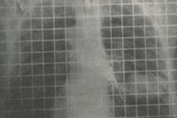
In 1929, Moniz's surgical colleague Reynaldo dos Santos, a professor of surgery in Lisbon, introduced percutaneous translumbar aortography (TLA) by direct aortic puncture with injection of a sodium iodide solution. Three years later, Santos described the technique (figs. 5 and 6). Fig. 7 shows abnormal vascularity in a sigmoid tumor; note the stationary grid lines.
 Fig. 4: Cerebral angiography (1932).
 Left, fig. 5: Dos Santos (1932). Right, fig. 6: Technique of aortography (1932).
 Fig. 7: Translumbar aortography for sigmoid tumor shows abnormal vascularity (1932).
TLA remained a standard for vascular imaging until the 1980s, when I was taught it as a trainee registrar at London's Hammersmith Hospital. Other members of Moniz's team ware equally innovative and successfully introduced pulmonary angiography (de Carvalho and Almeida Lima), lymphography (Monteiro), phlebography (Joäo Cid des Santos [son of Reynaldo]) and portal venography (Pereira).
In 1929, Werner Forssmann introduced a well-oiled ureteral catheter via an antecubital vein into his own right atrium. Two years later, Moniz, de Carvalho, and Almeida Lima used the Forssmann method to demonstrate the pulmonary vasculature with an injection of sodium iodide (fig. 8).
The Portuguese School introduced many aspects of clinical angiography in the 1930-1950 period, but the international adoption of their techniques was severely delayed by World War II.
 Fig. 8: Moniz and De Carvalho (1932) inscribed by Moniz: "Hommage de Egas Moniz."
The next major development related to the method of delivery of contrast medium into vessels and the heart chambers was achieved by Sven Seldinger in 1956, working at the Karolinska Clinic in Stockholm. He introduced the needle-guidewire catheter replacement technique that permits selective catheterization and injection of most vessels from a simple puncture. This technique is now routine.
Dr. Adrian Thomas is chairman of the International Society for the History of Radiology and honorary librarian at the British Institute of Radiology.
References
- Dos Santos R, Lamas AC, Caldas JP. Artériographie des Membres et de l'Aorte Abdominale. Paris: Masson et Cie, éditeurs; 1932.
- Moniz E, De Carvalho L. A visibilidade dos vasos pulmonares (Angiopneumografia). Lisboa: Imprensa Libanio da Silva; 1932.
- Moniz, E. Diagnostic des Tumeurs Cérébrales et épreuve de l'éncephalographie Artérielle. Paris: Masson et Cie, éditeurs; 1931.
- Veiga-Pires JA, Grainger RG, eds. Pioneers in Angiography: The Portuguese School of Angiography. Lancaster: MTP Press; 1982.
The comments and observations expressed herein do not necessarily reflect the opinions of AuntMinnieEurope.com, nor should they be construed as an endorsement or admonishment of any particular vendor, analyst, industry consultant, or consulting group.