U.K. researchers have developed a computer-based simulator for estimating radiation dose in computed radiography (CR) studies. The researchers believe their "virtual phantom" could prove more effective than physical phantoms by enabling clinicians to gauge the impact of patient anatomy when imaging parameters are changed.
In the U.K., chest x-rays constitute approximately 19.6% of all radiography exams. Because chest x-ray is performed so frequently, optimization of radiographic technique is important, wrote Craig Moore, a physicist and radiation protection adviser at Queen's Centre for Oncology and Haematology at Castle Hill Hospital in Cottingham, U.K., and colleagues in an article published online ahead of print in the British Journal of Radiology (17 January 2012).
Previous research has relied on physical phantoms to assess the impact of CR system noise on radiation dose when imaging parameters are changed. But physical phantoms don't simulate the most significant contributor to noise during imaging studies -- patient anatomy.
To develop a more accurate methodology, Moore and colleagues developed what they call a digitally reconstructed radiograph (DRR) -- a computer simulation of a 2D radiograph created from CT data. The model contains realistic patient anatomical noise produced at various equipment settings, such as tube potentials, receptor air kerma, and scatter rejection methods.
"There is guidance on optimum x-ray radiographic technique with film-screen systems for chest radiography, but none for digital imaging systems such as CR," Moore said in an interview with AuntMinnieEurope.com. "Therefore, research must be carried out to determine optimum radiographic techniques (e.g., tube voltage [kVp], tube current-time product [mAs], filtration, scatter rejection methods) for CR imaging modalities."
They tested the model in a "virtual clinical trial" in which four experienced image evaluators graded images of average and obese adult patients at different potentials, receptor doses, and scatter rejection techniques on an Agfa HealthCare CR system using MD-4.0 phosphor plates. The quality of the images was evaluated using visually graded analysis. The researchers also assessed the influence of rib contrast.

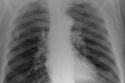
 Simulated chest images of an average-sized patient reconstructed at 50 kVp (a) and 150 kVp (b). Image (a) is of higher contrast, which is expected. All images courtesy of Craig Moore.
For average-sized patients, image quality improved when tube potential was reduced compared with the reference (102 kVp) and no scatter rejection was indicated. For obese patients, it has been shown that an antiscatter grid is indicated and should be used in conjunction with as low a tube potential as possible (while allowing exposure times, 20 msec), the researchers wrote. It is also possible to reduce receptor air kerma by 50% without adversely influencing image quality. They also found rib contrast did not interfere at any tube potential.
In 50 average patients who were reconstructed without scatter rejection, readers gave higher marks for images acquired with lower tube potential (kVp) settings, demonstrating that image quality improves with lower kVp, the researchers said. Visual grading analysis system scores ranged from 0.41 for 50 kVp to 0.03 for 109 kVp, but there was very little difference between image quality at tube potentials greater than 102 kVp (0.03 at 109 kVp to 0.07 at 150 kVp), according to the authors.

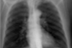
 Simulated chest images of an average-sized patient reconstructed with an antiscatter grid (a) and without a grid (b). Image (a) provides greater detail, especially in the denser regions of the chest, which is expected.
"Although there is a trend toward increased image quality at low tube potentials, it was only possible to statistically distinguish between 50 and 90 kVp, 80 and 90 kVp, and 80 and 109 kVp," Moore and colleagues wrote. "All other combinations demonstrated p-values > 0.102. Nevertheless, these results show that for matched effective dose, image quality improves with lower tube potentials, which is similar to results reported in the literature."
However, to compensate for the reduced intensity of the x-ray photons, an increase in mAs would be needed, they noted.
For average adult patients, low tube potentials (102 kVp) and no scatter rejection provide superior image quality, according to the researchers.
"However, it has been shown that for obese adult patients an oscillating focused antiscatter grid is indicated, and should be used in conjunction with as low a tube potential as possible, as this combination provides superior image quality to any other investigated during this study," they stated.
Measurements with a chest phantom on a clinical x-ray system demonstrate any tube potential used without a grid can be used for average patients to satisfy the 20-msec exposure time limit, but at least 90 kVp must be used for obese patients, the authors concluded. It has also been shown that receptor air kerma through the lung region can be reduced to 1.5 µGy while maintaining an adequate level of image quality, and the influence of rib contrast interfering with image evaluation is minimal.