As the use of radiography and imaging devices increases in the operating room, so does the need for radiation-related training. Technological improvements to a cutting-edge C-arm radiation awareness and protection training module can allow more accurate simulation of scattered radiation. This will help ensure that operating room personnel are better equipped to prevent unnecessary exposure to both themselves and patients.
Radiation exposure awareness and protection is obligatory for C-arm operators under German law. Developed by three German institutions -- the University of Applied Sciences and Arts in Hannover, the Medical University of Göttingen, and the Technical University of Brunswick -- virtX was first introduced as an interactive radiation-free, hands-on simulation approach to radiation education in 2005.
To date, radiation diffusion has been difficult to compute. Now the award-winning training module is set to include a more realistic simulation of scattered radiation using the Geometry and Tracking (GEANT4) toolkit. It employs Monte Carlo methods to simulate the passage of particles through matter.
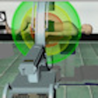
"Last summer we only had a very simple, ball-like visualization that abstracts intensively from the physically correct distribution of scattered radiation, but now we are using a new physical simulation module in combination with massive parallel computing on modern graphic card hardware," said Oliver Bott, PhD, a computer scientist at Hannover's University of Applied Sciences.
This new technology facilitates simulation of correct distribution near real-time. The trainee can instantly understand the distribution of scattered radiation in the OR and the immediate effects of radiation protection measures such as modifying apertures, distance to radiation source, or position of the image intensifier.
With virtX, C-arm systems fitted with an electromagnetic motion-tracking device feed information to the computer about the location of the C-arm in relation to a dummy patient. The computer then uses the position of the C-arm to compute a model radiograph from a store of CT images, while an embedded scattered radiation simulation system measures the resulting "virtual" radiation extension so that the surgeon can take steps to reduce it.
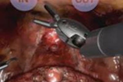
 The user interface shows visual and interactive learning elements of image quality and radiation output. All images provided courtesy of Oliver Bott, PhD.
"Nurses, surgeons, and technicians need to know how to either operate these systems or know which adjustments or positioning is necessary to get a certain image needed to assess a specific surgical situation," Bott said. "Here we have a tool that directly improves training by giving immediate feedback to a trainee's handling of a C-arm."
Achieving a better spatial understanding, as well as memorizing the relevant adjustments for different standard views, is vital to avoid producing unnecessary radiation, as can happen while views are adjusted for correct visualization.
Despite a long history of training and educational activities focusing on improving awareness, there is a substantial lack of understanding about the dangers of radiation exposure, especially with scattered radiation, according to Bott. This may be due to the theoretical nature of current training programs for radiation protection, he added. Until now, courses on radiation protection have used fixed pictures and precalculated videos. It is hoped that new e-Learning modules based on computer simulations will improve education, beyond the practice of C-arm operation, through interactive visual and practical elements.
The simulation-based approach of virtX could also be adopted for 3D C-arms, dental radiography, or bucky tables. Current development is focused on the integration of specific surgical procedures that make intensive use of C-arm technology, such as femoral nailing.
"Because it is difficult to combine hands-on training courses with real devices and patients, especially for minimally invasive techniques, e-Learning approaches can be a valuable alternative," Bott said.
 Surgeons in Davos, Switzerland, get to grips with the PC version of the virtX training package. Oliver Bott is seated on the left.
Both virtX and its sibling virtusMED, aimed at ultrasound training for medical students, can exist as a PC software package for training either at a center or at home using just a keyboard and mouse. A bigger package aimed at institutions combines the software with electromagnetic trackers and real imaging systems. Now commercially available, neither of these latter formats is cheap, but evidence of their added value is emerging. A study published in 2008 that described the use of virtX combined with a C-arm compared with traditional training showed that the former had a significant effect on the time needed to obtain a high-quality image with optimal radiation exposure protection (Methods of Information in Medicine, 2008, Vol. 47:3, pp. 270-278). Also, the creators of the package won a Summa Cum Laude award for their poster presentation about their research at the RSNA 2009 meeting.
"Based on proven benefits, e-Learning approaches are a useful addition to current course structures, especially in radiology education, including interventional training," Bott said.