European researchers found no statistically significant benefit in the use of dual-energy digital radiography (DR) compared with conventional DR for detecting pulmonary nodules. The predominance of small lung nodules in the multicenter study may have exceeded the ability of dual-energy DR to detect them, they wrote.
Radiography's role in chest imaging has been eclipsed by CT, due to the latter modality's ability to visualize small lung nodules that are malignant or at risk of becoming malignant. This enables clinicians to catch lung cancers at a stage when they can be treated more effectively.
However, the increasing use and flexibility of DR has raised the prospect that digital technologies might help radiography gain an edge over CT. To this end, investigators have been pursuing everything from computer-aided detection (CAD) to tomosynthesis to dual-energy subtraction in an effort to unlock DR's potential.
In a dual-energy study, two x-ray exams are taken, each at a different energy level. Because the x-ray attenuation of bone and soft tissue varies depending on the energy level of the x-ray beam, either bone or soft tissue can be eliminated from the final image, removing overlapping structures and enabling clinicians to focus on their area of interest.
But does dual-energy subtraction translate into improved detection of malignant lung nodules? Researchers from Europe sought to answer this question in a prospective study that compared dual-energy DR with conventional DR. Their results were published in the April 23, 2008, online edition of European Radiology.
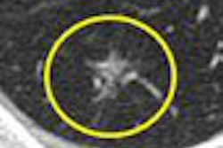
Researchers from four medical centers examined 100 patients with a total of 140 CT-confirmed lung nodules between April 2003 and April 2005. The patient population had a median age of 64 years (range 22-92 years) and consisted of 47 men and 53 women.
Patient enrollment was limited to individuals with no more than six lung nodules with a size of 3-45 mm (median size 11 mm), and patients were excluded for severe diffuse pulmonary disease such as pneumonia or idiopathic pulmonary fibrosis. Seventy-one patients had known malignant disease, 18 had no evidence of malignancy, and the status of pulmonary nodules in the remaining 11 patients was unconfirmed.
Patients were examined on a flat-panel DR system based on a cesium iodide scintillator and amorphous silicon photodiode transistor array (Revolution XQ/i, GE Healthcare, Chalfont St. Giles, U.K.). The dual-energy exam consisted of standard posteroanterior (PA) radiographs, as well as subtracted soft-tissue and bone images, the authors wrote.
The dual-energy study was conducted with one exam at 120 kV and 2 mAs at an equivalent film speed of 400, while the second study was performed at 60 kV and 8 mAs with an equivalent film speed of 1,000. Postprocessing algorithms were also applied to the data.
Images were independently reviewed by five radiologists from the four medical centers; all readers were general radiologists with experience in chest radiography. Readers reviewed the images on soft-copy PACS workstations.
Cumulative results of the study for all five readers are indicated below:
|
The marginally higher marks for dual-energy DR in the study were not statistically significant, the authors reported. Moreover, there were no statistically significant differences in reader performance when measured by area under the curve (Az) values.
The readers noted that their results differed from previous studies, pointing to a positive, statistically significant effect of dual-energy DR on lung nodule detection. The study's focus on smaller lung lesions could have affected the results, they wrote, with almost half (47.7%) of the nodules in the study being smaller than 10 mm.
"As expected, the majority of nodules missed by the reviewers were those of the smallest sizes and seem to have been too small to take advantage of the (dual-energy) technique," the researchers wrote. "Results of a subgroup analysis for different lesion sizes revealed significance for nodules of 20 to 29 mm only and may be coincidental."
On the positive side, the researchers said that the technique, with its dual exposures, did not seem to add significantly to the total radiation dose delivered during the exam. Total radiation dose, including PA and lateral shots, increased 14% by applying the dual-energy mode versus standard chest DR.
By Brian Casey
AuntMinnie.com staff writer
May 1, 2008
Related Reading
CAD provides mixed benefits for DR lung exams, March 8, 2008
DR pumps new life into tomosynthesis-based radiography, October 29, 2007
Dual-energy digital x-ray still looking for acceptance, February 22, 2007
Copyright © 2008 AuntMinnie.com