MRI plays an important role in the early diagnosis and management of tubal pregnancy, and certain features on MRI such as "three rings" make it ideal for early diagnosis of this condition, according to an article published in European Radiology.
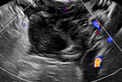
Researchers reviewed the medical records, ultrasound scans, and MRI results of 27 patients who were clinically suspected of having ectopic pregnancy. The research team, led by Dr. Ming-Jue Si from the radiology department at No. 9 People's Hospital in Shanghai, found ectopic pregnancy does indeed exhibit certain features on MRI, including a gestational sac-like structure with a "three rings" appearance on T2-weighted images, presence of solid components in the sac, dilatation of the affected tube with hematosalpinx, and tubal wall enhancement.
"MRI plays an important role in the early diagnosis of tubal pregnancy and provides accurate evaluation of the lesion to help make management decisions," the study authors wrote (Eur Radiol, 15 September 2015). "Radiologists should choose the most necessary MRI sequences according to the requirements."
What is ectopic pregnancy?
Ectopic pregnancy is defined as the implantation of a fertilized ovum outside the uterine cavity, and it accounts for approximately 2% of all pregnancies. The most common location is the fallopian tube, which accounts for 97% of the cases. Ectopic pregnancy is a life-threatening surgical emergency and a leading cause of maternal mortality during the first trimester of pregnancy.
Mortality has dropped in the past two decades due to advancements in early diagnosis and treatment. Until recently, most ectopic pregnancy diagnoses have been based on pelvic ultrasound along with quantitative measurement of β-human chorionic gonadotropin (β-hCG). However, if the gestational sac is small without embryocardia beats, a mixed extrauterine mass is detected via ultrasound, which is inconclusive for ectopic pregnancy diagnosis, according to Si and colleagues.
If the patient is in the early stages of ectopic pregnancy and requires conservative treatment, a detailed evaluation of the gestational sac and the affected fallopian tube is necessary to help clinicians make proper management decisions.
This is where MRI comes in. The modality is increasingly being used as a problem-solving adjunct to ultrasound, as it provides additional information for complicated cases.
To date, only a few studies focusing on MRI for ectopic pregnancy have been conducted, and most were limited to case reports or parts of a review series in which the characteristic MRI features were not extensively discussed.
Si's team sought to rectify this by determining the role of MRI in the early diagnosis and management of ectopic pregnancy and also investigating the characteristic MRI features with respect to the pathological findings.
The results
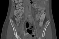
The researchers retrospectively reviewed 27 tubal pregnancy cases -- both clinical and MRI features -- on a 1.5-tesla magnet (Signa, GE Healthcare).
They found a thick-walled gestational sac-like structure was demonstrated laterally to the uterus in all cases. On T2-weighted images, the thick wall typically exhibited three separate rings in 22 cases (81%), among which 17 cases (63%) displayed small vessels and six cases (33%) exhibited small areas of fresh hemorrhage inside the thick wall.
Also, the sac contents demonstrated nonspecific liquid in 26% of the cases, papillary solid components in 56%, and fresh blood or fluid-fluid level in 19% of the cases.
Dilatation of the affected fallopian tube associated with hematosalpinx was demonstrated in 18 cases (67%), and marked enhancement of the tubal wall was observed in 22 cases (81%). No correlation was found between the size of the gestational sac and the estimated gestational age.
Characteristics and limitations
The most characteristic MRI feature of ectopic pregnancy is the cystic lesion with a thick wall typically exhibiting a "three rings" appearance on T2-weighted images with or without solid components, which is rarely observed in other adnexal masses and, thus, can be used as a key imaging feature, according to the researchers.
The pathological examination showed the thick middle ring was composed of chorionic villi tissue, which contained abundant fetal capillaries and maternal blood in the interstitium, thereby resulting in hypersignal intensity on T2-weighted images and marked enhancement after administration of contrast material. The thin outer ring was formed by the adjacent tubal wall, whereas the thin inner ring was formed by extraembryonic coelom and amnion without blood vessels, thus resulting in hyposignal intensities on T2-weighted images, the researchers added.
The study is not without its limitations, namely the retrospective nature and small sample size. Also, 89% of the cases were associated with an unruptured or slightly ruptured ectopic pregnancy, and the MRI features of completely ruptured ectopic pregnancies were not fully discussed.
"This condition is a life-threatening surgical emergency and is difficult to manage for a long-time examination like MRI," the study authors wrote. "The suspicion of tubal rupture more often depends on the clinical settings, including acute severe abdominal pain, guarding and rebound tenderness at physical examination, hemodynamic instability, and dropping hematocrit."
Lastly, ectopic pregnancy frequently coexists with a corpus luteum cyst with differences in origination and morphology. However, when the lesions are tiny, it can be very difficult to distinguish between them by MRI.