Experience is not a factor when it comes to interpreting mammograms on lower resolution, cheaper viewing devices. And LCD monitors with image manipulation software are suitable for viewing mammograms for training purposes, found a U.K. study that included findings on iPhone use.
"We expected the clinical monitors to very clearly be superior," noted lead author Yan Chen, PhD, from the department of computer science at Loughborough University, in an email to AuntMinnieEurope.com. "Surprisingly, the more typical 'office' monitor fared much better than we expected."
Also, one study participant using an iPhone correctly identified all the small microcalcification clusters on the iPhone image, which suggests the pan/zoom function implemented in the Osirix software on the iPhone was capable of displaying many of the small mammographic features. However, it is difficult for a reader to ensure the whole image has been completely viewed when panning and zooming on such a small display.


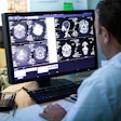
Examples of participants examining images on the three different displays. The ambient lighting levels were altered for photographic purposes. Images courtesy of Yan Chen, PhD.
"Poor performance on the lowest resolution screen probably relates to a combination of factors with participants not being able to navigate appropriately around the zoomed image to ensure that all parts of the image have been properly assessed as well as the 'pixilation' phenomenon making subtle features uninterpretable," the researchers wrote in their online article in European Radiology (3 June 2015).
With practice, other iPhone users might achieve the same results as the reader who picked out all microcalcifications, Chen stated in her email.
"Bear in mind that we deliberately used an early iPhone with lower resolution than current iPhone models to see just what could be achieved with such low-resolution displays," she noted. "One iPhone user was particularly good at identifying all microcalcifications as she took a very patient approach and used image manipulation very well. This showed that it could be used successfully with these particular test mammograms; however, it took a long time and hence was not very popular with readers."
Such a small display is not ideal, however. Better interaction techniques and improved image manipulation software would be useful -- this is one avenue of the group's current research. "It is important for research to examine the limits of the use of such devices for training purposes," Chen added.
Interpretation challenges
Digital mammograms pose a particular challenge for interpretation because matrix sizes are often in excess of 4,800 x 6,400 pixels, compared with CT images that are often 512 x 512 pixels for a single slice. Other important factors are contrast ratio and grayscale depth.
Right now, high-resolution dual-monitor PACS workstations with 5-megapixel (MP) resolution and 12-bit grayscale display are considered the cream of the crop, and, as such, they are expensive, with limited availability in many departments. The availability and cost of high-resolution workstations has implications for delivering reader training and education, according to Chen and colleagues.
"Lower resolution monitors such as a standard office PC monitor or handheld computing devices are significantly cheaper and widely available," they wrote. "Little is known about the adequacy of lower resolution viewing devices in delivering teaching and training of mammographic interpretation."
The researchers sought to determine if mammographic interpretation was possible by combining less sophisticated viewers with appropriate image manipulation software such as zoom, pan, and window level/width adjustment, and to compare performance with a standard high-resolution PACS workstation.
On three occasions over eight months, 14 consultant (senior) radiologists and reporting radiographers read 40 digital mammography screening cases on three different displays: a digital mammography workstation, a standard LCD monitor with a 1.8-MP display, and a smartphone with a 0.15-MP display. Standard image manipulation software was available for use on all three devices.
The researchers used receiver operating characteristic (ROC) analysis and analysis of variance (ANOVA) to determine the significance of differences in performance between the viewing devices with/without the application of image manipulation software. They also assessed whether the reader's experience affected performance.
Performance was significantly higher (p < 0.05) on the mammography workstation compared with the other two viewing devices. When image manipulation software was applied to images viewed on the standard LCD monitor, however, performance improved to mirror levels seen on the mammography workstation with no significant difference between the two. Image interpretation on the smartphone didn't fare quite so well -- it was uniformly poor minus the one reader.
Reader experience had no significant effect on performance across all three viewing devices, the researchers explained. However, as anticipated, the more experienced readers performed better on all three devices.
Potential cost savings
The results of the study suggest facilities might be able to save themselves some money by using LCD monitors with image manipulation software instead of opting for the high-resolution monitors -- something Chen calls "exciting." But she is quick to note that the LCD monitors could be used for certain circumstances but not others.
"Facilities could save money by using normal monitors in some circumstances, for example, for mammographic interpretation training," she wrote. "It is important to note that we are not saying that subclinical mammographic monitors could be used for clinical reporting of mammograms such as in breast screening."
For training purposes though? Using less expensive monitors means training opportunities can be extended beyond the radiological reporting room itself, Chen said.
"Not every aspect of mammographic interpretation could be learnt using such subclinical displays, but a lot could be achieved," she added.
Other research plans include eye tracking -- monitoring just how radiologists examine mammographic images in different conditions. Chen and colleagues'' research is part of an ongoing national PERFORMS scheme in which every year they assess the skills of the U.K. breast screening workforce on test sets of difficult mammograms.


