Accurate alignment of two medical images in which a particular structure is present in only one of them poses a serious challenge for registration algorithms. To address this problem, researchers from University Medical Center Utrecht in the Netherlands have proposed the addition of a geometrical penalty term into a conventional nonrigid registration framework. They have demonstrated their approach using an example of cervical brachytherapy MR images.
Cervical cancer is often treated using a combination of external-beam radiotherapy and brachytherapy, with MRI employed for target delineation prior to each treatment. Accurate registration of a patient's different pretreatment images should enable better estimation of the cumulative delivered radiation dose. But registration is hindered by the presence of the large brachytherapy applicator (which will also alter the geometry of surrounding tissue) in only one of the images.
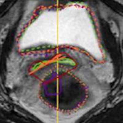
 Transversal (top) and sagittal (bottom) view of a moving image overlaid with the propagated delineations of the bladder, applicator, and rectum. Green lines denote the gold standard delineations. Propagated delineations of conventional registration with no applicator mask (CnM), conventional registration with an applicator mask (CwM), and registration including the proposed MSP term (MSP) are shown in purple, red, and orange, respectively.
In deformable image registration, a so-called moving image (in this case, the external-beam MRI) is warped to the fixed image (the applicator-containing brachytherapy MRI). The method proposed by the Utrecht team exploits the fact that the missing structure can be considered to have zero volume after mapping it to the external-beam image. By introducing a geometrical penalty term that minimizes the volume of the void left by the applicator's removal, this mapping can be established (Physics in Medicine and Biology, 2 July 2014, Vol. 59:15, pp. 4033-4045).
"While this study considers cervical brachytherapy, this is a general problem in registration," explained Floris Berendsen, a PhD candidate on image registration at the Image Sciences Institutein Utrecht. "I am collaborating a lot with our radiotherapy department -- it is a good source of data, and tough and relevant registration problems."
Data analysis
Berendsen and colleagues incorporated their proposed missing structure penalty (MSP) term into the general purpose nonrigid registration framework from the elastix toolbox. They tested the resulting algorithm on 18 pairs of cervical MRI scans -- each pair constituting a fixed image with the applicator in place and a moving image without an applicator.
They used two sets of data from five patients (study sets A and B) to develop the method, and a third data set from eight patients for evaluation. In study set A, moving images were acquired immediately after applicator removal; in the other two sets, images were obtained the week before brachytherapy, during planning of the external-beam fraction.
The researchers evaluated three types of registration: conventional registration with an applicator mask (in which masked voxels in the external-beam image do not influence registration); conventional registration without an applicator mask; and registration including the proposed MSP term. The mapping established by each was then used to transform all manual delineations of the fixed image to the moving image.
These transformed delineations (U') were then compared with gold-standard manual delineations (Ugold) of the bladder and rectum (organs-at-risk), and the surface of the void (a combination of vaginal and uterine walls) in the external-beam image. The applicator was also manually delineated for use in the proposed MSP term and the mask-based registration.
Registration accuracy was assessed by measuring surface distances of the closest plane or point on U' for all points on Ugold, and vice versa. The researchers combined these distances and characterized them in terms of maximum, 90th percentile and median distances. Compared with conventional registration (with or without the mask), use of the MSP term improved alignment accuracy for the void, with average values of the above three measures substantially improved for all datasets. In two of the three datasets, alignment of structures near to the missing applicator was also improved.
The team also determined the applicator residual volume (in the external-beam image) for the three registrations. The ability of the penalty term to minimize the void volume was shown by the very low average residual volumes obtained (0-2 mm3) compared with those from conventional registrations (12-42 mm3). Without the penalty term, the registration process could not find a mapping with a minimum applicator void volume.
"In brachytherapy image registration, the penalty term substantially improves the alignment of the missing applicator structure to the gold-standard anatomical position," the researchers concluded.
Berendsen is now working on a related registration problem, in which the surfaces of organs have slid along each other in between two scans.
© IOP Publishing Limited. Republished with permission from medicalphysicsweb, a community website covering fundamental research and emerging technologies in medical imaging and radiation therapy.