Move over mammography: There's a new imaging device to detect breast cancer. Or there will be in about four years if everything goes according to plan with a large Dutch-led research consortium funded by the European Union (EU).
The University of Twente in the Netherlands and partners in a large consortium called Pammoth are due to receive grants of more than 5 million euros, predominantly from the EU, to develop a device that uses photoacoustic and ultrasound imaging. It will also combine images generated by both techniques, and the project leaders expect a prototype will be ready for large-scale testing and production in four years.
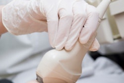
"The proposed device is expected to shorten the time to accurate diagnosis substantially," the university said in a release. "It will also be suitable for younger women, where x-ray mammography usually does not perform well. The device does not use radiation or contrast media, which are potentially harmful, and does not cause any pain to the woman being examined."
Device specifics
It is well-known mammography has its shortcomings: It doesn't pick up every tumor and is particularly inadequate in women with dense breasts. For years, researchers have worked to combat this problem or sidestep it by adding adjuncts, namely MRI and ultrasound. MRI produces excellent image quality but can be expensive and time-consuming depending on the chosen protocol. Ultrasound is also beneficial but is often operator-dependent and thus not as reliable. Enter photoacoustic mammography, which project lead Dr. Srirang Manohar calls "a dream imager."
 Dr. Srirang Manohar. Photograph by Robert Molenaar.
In photoacoustic mammography, the breast is illuminated with laser light. This laser light is converted into ultrasound in regions where higher concentrations of blood are present, such as areas around malignant tumors, according to the university. The ultrasound produced travels from the tumor to the surface of the skin.
Manohar and colleagues will improve the technique and introduce functionalities such as various colors of laser light to enhance visualization of the tumor, as well as to yield information about the oxygen saturation of the blood in tumors, indicating whether they are malignant or benign. However, photoacoustics alone cannot provide anatomic information, which is why they are also including ultrasound imaging, he told AuntMinnieEurope.com.
The ultrasound portion of the Pammoth imager will yield a 3D image, with the ultrasound waves being generated in an unconventional manner, the university said. And, unlike regular ultrasound, handheld scanners will not be used, eliminating operator dependence.
"The images from the two systems -- photoacoustic and ultrasound -- will be combined together," Manohar said in the university's release. "This will result in simultaneous 3D information about disease-specific optical contrast, as well as about the ultrasound properties, which provide anatomic information within the breast. Further, we wish to do this combined imaging in real-time."
The researchers plan to start the project on 1 January with a kickoff meeting that will include clinical advisers and patient advocacy representatives. They'll take the first steps toward finalizing the specifications of the instrument.
"From then on, the work starts in earnest," he said in an email.
The project also extends beyond breast cancer -- Manohar expects it to lead to various improved subsystems for data acquisition and ultrasound detection and a new laser.
Who's involved?
Manohar, from the University of Twente's Biomedical Photonic Imaging Group, heads the consortium, focusing on photoacoustic imaging and research for generating ultrasound. PA Imaging, a University of Twente spin-off company, will develop rapid, low-noise electronics for the detectors and will ensure that the various partners' components are integrated into the functioning clinical prototype, the university said.
Medical Spectrum Twente along with the University of Twente will conduct a pilot study to test the performance of the system by imaging the most common breast tumors once the prototype is complete. Researchers from University College London will work on image reconstruction methods, and will be responsible for the mathematics and calculations required to compute the images, the university added.
The developed mathematics will be converted into usable and rapid algorithms to enable real-time imaging at the Brno University of Technology, Czech Republic. Scientists from the University of Bern in Switzerland will have a role in the analysis of ultrasound images and will develop specialized breast phantoms.
The French company Imasonic will design highly sensitive, multielement detectors for detecting the ultrasound, the Lithuanian firm Ekspla will develop the lasers needed for the system, and the German company TP21 will be responsible for managing the project and the internal information infrastructure.
The project is financed by the EU's Horizon 2020 program to the tune of 4.35 million euros. The Swiss government is providing 750,000 euros.