X-ray mammography, the current gold standard for breast cancer detection, has an average false-negative rate of 20%, or higher in women with dense breast tissue. Diffuse optical imaging (DOI) has been proposed as a nonionizing and relatively inexpensive alternative method for detecting breast cancers, including those in dense tissues. Now, U.S. scientists have developed a handheld optical scanner with the potential to image breast cancer in real-time.
The device, developed primarily at Florida International University, uses a near-infrared laser diode to produce an image of the breast tissues. The images are created by mapping the optical absorption at 785 nm -- the point at which the absorption spectra of oxy- and deoxyhemoglobin intersect -- and thus provide an indication of total hemoglobin concentration. Increased hemoglobin concentration is typically observed at the tumor site due to tumor angiogenesis (Biomedical Physics & Engineering Express, December 2015, Vol. 1:4).


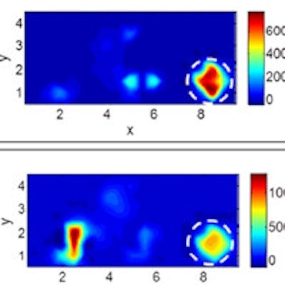
X-ray, ultrasound, and diffuse optical images of the left breast from a 51-year-old breast cancer patient. (A, B) X-ray mammography images; the yellow arrows indicate three lesions in the breast. (C, D) Ultrasound images showing two malignant (L1, L2) and one benign lesion (L3). (E-H) Diffuse optical images. The schematics (not to scale) indicate the approximate location of the tumors (red circles) and the probe (blue boxes). The white dotted circles in the diffuse optical images indicate a rough estimate of the tumor locations; the color bar indicates absorption due hemoglobin.
The researchers used the handheld optical imager to image five women diagnosed with breast cancer prior to surgical intervention. They recorded images at various locations on both the tumor-containing and contralateral breasts. The images revealed regions of higher intensity (indicative of increased vasculature and higher blood content) at the tumor location. A preliminary result also indicated the potential to image lymphatic spread.
One advantage of the new device is that it is more adaptable to breast shape and density, and it allows imaging of the chest wall regions, which are harder to image with conventional techniques. "The women scanned always commented on how comfortable it was to be scanned by our device -- many of them said that they didn't feel anything," explained Sarah Erickson-Bhatt, PhD, an author on the paper.
"Eventually, we hope that physicians will be able to use this for real-time imaging of breast tissues as part of regular visits by the patients," added Anuradha Godavarty, PhD, also an author of the paper. "We're currently working on the mathematical tools required to process the images and produce 3D tomographic images, in order to determine tumor size and depth."
The researchers' ongoing efforts involve extensive clinical work to demonstrate the capability of the device to prescreen for any breast abnormality, followed by seeking U.S. Food and Drug Administration (FDA) approvals prior to clinical use.
© IOP Publishing Limited. Republished with permission from medicalphysicsweb, a community website covering fundamental research and emerging technologies in medical imaging and radiation therapy.