A new breast elastography technique is demonstrating promise as a reliable method for measuring the stiffness of healthy breast tissue. Standard values proposed by the German researchers may even differentiate parenchyma and fatty tissue from breast lesions.
In a study evaluating a new shear-wave elastography technique developed by Siemens Healthcare called virtual touch tissue imaging quantification (VTIQ), researchers from the University of Heidelberg found that the method yielded significantly higher values for parenchyma than fatty tissue. It also showed good retest reliability (European Journal of Radiology, 7 August 2013).
While early elastographic methods relied on manual compression and decompression applied by the examiner, VTIQ automates the tissue compression process to improve examiner independence and reproducibility, according to Dr. Michael Golatta, from the breast unit at the University of Heidelberg in Germany, and colleagues. The probe then generates a longitudinal push pulse that causes minimal localized displacement and is tracked by a detection pulse.
Because the speed of the shear waves propagating through the tissue is proportional to the stiffness of the tissue, a colored map in the region of interest (ROI) gives information on the tissue stiffness in the ROI, according to the authors.
VTIQ comes into play for differentiating benign and malignant lesions. To differentiate the two, it is necessary to describe and analyze individual breast tissue composition, namely breast parenchyma and fatty tissue.
However, few studies have evaluated the elastographic behavior of regular breast parenchyma and fatty tissue, and only one of those evaluated the VTIQ technique in a prerelease version, according to the researchers. As a result, they sought to prospectively explore breast parenchyma and fatty tissue stiffness and evaluate the retest reliability with the new method of VTIQ.
B-mode ultrasound and VTIQ were performed in 132 breasts in 97 women. Mean values of VTIQ for parenchyma and fatty tissue were compared between those measured in healthy breasts and the surrounding of histologically proven benign and malignant breast lesions. The researchers also reviewed VTIQ values according to breast density measured by the American College of Radiology (ACR) categories.
In 132 breasts, the mean VTIQ values in parenchyma were significantly higher than in fatty tissue (3.23 m/sec ± 0.74 versus 2.5 m/sec ± 0.61; p < 0.0001). In healthy breasts as well as in the surrounding of benign or malignant lesions, the VTIQ values of parenchyma were similar (p = 0.12). In fatty tissue, small differences between mean VTIQ values of 2.25 m/sec ± 0.51, 2.52 m/sec ± 0.48 and 2.65 m/sec ± 0.71 (p = 0.01) in the respective groups were observed.
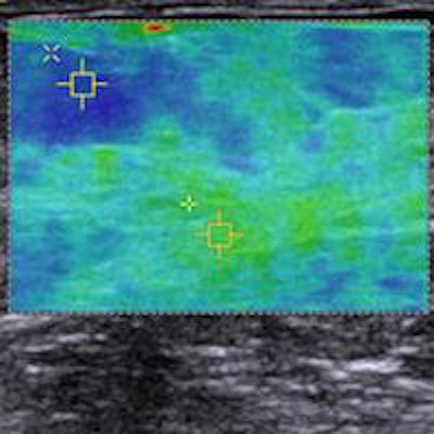
 VTIQ image of a healthy breast showing fatty tissue (1.38 m/sec) and parenchyma (3.33 m/sec). Image courtesy of Dr. Michael Golatta.
The comparison of mean VTIQ values of parenchyma and fatty tissue in more and less dense breasts (ACR 1 + 2 versus ACR 3 + 4 breasts) yielded no statistically significant difference, the researchers found.
The researchers proposed standard values of VTIQ for healthy breast tissue: Parenchyma showed a mean of 3.23 m/sec (31.3 kPa) with a standard deviation of 0.74 m/sec (1.6 kPa) compared with 2.5 m/sec (18.8 kPa) for fatty tissue with a standard deviation of 0.61 m/sec (1.1 kPa). The difference between the two was statistically significant (p < 0.0001).
"Even though our values for breast parenchyma and fatty tissue show an overlap with benign breast lesions, there is a significant difference to the, in general, stiffer malignant lesions," they wrote. "Dichotomizing the cohort by the density of the breast tissue into ACR 1 + 2 and ACR 3 + 4 showed that there is no difference in the stiffness of breast tissue depending on breast density."
In addition, comparing the mean stiffness of parenchyma and fatty tissue in the surrounding of benign, malignant, or normal breast tissue revealed no relevant differences, they added.
The method's retest reliability, as assessed with three independent measurements, was moderate, yielding an interclass correlation of 0.52 (p < 0.0001).
"Based on this result, VTIQ is a reliable method that can be used for breast diagnostics," the researchers concluded. "The proposed standard values may ... differentiate parenchyma and fatty tissue from breast lesions. Stiffness of healthy breast tissue seems to be robust with respect to breast density and surrounding tissue."
A second VTIQ paper is currently in the review process and will hopefully be published soon, Golatta wrote in an email to AuntMinnieEurope.com.