Arguably one of the most eye-catching and unexpected moments at last week's RSNA meeting was the sight of Willi Kalender, PhD, and Dr. Christiane Kuhl enjoying a long, amicable chat in the Lakeside Center. These two well-known scientists have championed CT and MRI respectively for many years, fighting their corner with intense passion and evangelical-like zeal, but now they're forgetting their differences and working together on potentially ground-breaking research.
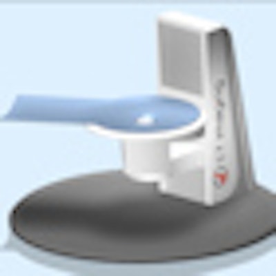
Kalender has developed a new CT system that he thinks will offer a significantly more accurate diagnosis of breast cancer for women with dense breasts, among others, through high contrast, 3D images. An examination on the scanner, which was displayed in Chicago at a special German Federal Ministry for Education and Research (BMBF) booth, will involve no more radiation exposure than standard screening mammograms.
 Sketch of breast CT scanner. The patient is positioned prone, and only one breast is exposed and examined at a time. All images courtesy of Willi Kalender, PhD.
Clinical trials of the device are due to begin in mid-2013, and will take place at the Friedrich-Alexander-University of Erlangen-Nuremberg in Germany, where Kalender is professor and chairman of the Institute of Medical Physics, and at the University of Aachen, where Kuhl is director of diagnostic and interventional radiology.
Kalender admits he has deep respect for Kuhl. "I know that she's the ideal person to come to an objective, scientific opinion about breast CT, particularly given her background in breast MRI," he told AuntMinnieEurope.com. "If I am able to convince her about the benefits of the system, then I'm sure I can convince anybody!"
The initial pilot study is likely to involve 30 patients with known breast cancer, and it will also include mammography and tomosynthesis examinations. A grant proposal is in preparation for submission to the German Research Foundation (Deutsche Forschungsgemeinschaft, DFG) in Bonn, and Kalender hopes ethical approval will be obtained soon. The research will be run by the Institute of Medical Physics, headed by Kalender, and supported by the Erlangen-based start-up company CT Imaging GmbH, of which Kalender is the founder, shareholder, and CEO. The goal is to present the first clinical cases at RSNA 2013, and to have 10 worldwide installations by the end of 2014.
At his RSNA refresher course on 30 November, Kalender explained that the gantry will provide comfortable patient-positioning with coverage of the full breast and the axilla, variable table height (about 70-170 cm), sequential and spiral scanning (25 cm in 10 seconds), and easy access to the patient for biopsy and therapy. From the system, he expects good detectability of microcalcifications of 100-150-micron diameter and soft-tissue lesions of 2 mm diameter, at an average glandular dose (AGD) of 2 to 4 mGy.
"We also offer dynamic contrast-enhanced scanning for improved analysis of differential uptake," he said. "Our objective is a 'one-stop shopping' modality for complete diagnostic workup on one device."
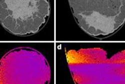
 Phantoms used to assess the performance of breast CT. Top left: Homogeneous 14-cm diameter phantom with soft-tissue lesions and areas of microcalcifications. Top right: Magnified portion of left image with the structures labeled. Bottom images show image details with window settings for soft tissue (left) and for calcifications (right).
In an article published by European Radiology in January 2012, Kalender and colleagues found that by micro-CT imaging of specimens, CT with high isotropic spatial resolution can provide the resolution for excellent assessment of microcalcifications with a signal above 5,000 HU at 60 kV. Using a dedicated breast CT system with cadmium telluride (CdTe) crystals and photon-counting electronics with 100-micron detector-element size for close to 100% dose efficiency, they assessed the potential for imaging microcalcifications of 100 microns to 200 microns in diameter, and soft-tissue lesions between 1 mm to 5 mm in diameter, by simulations at dose levels between 1 mGy and 6 mGy.
They clearly detected microcalcifications of 150 microns and soft-tissue lesions of 2 mm in diameter at an average glandular dose of 3 mGy. Separate displays are required for high-resolution microcalcification and for low-resolution soft-tissue analysis. In addition, total CT data acquisition time was below 10 seconds. Due to the high calcium signal at 60 kV, the existence of microcalcification clusters is clearly recognizable even when they are not resolved as single entities, according to Kalender.
At the end of the his RSNA course, he acknowledged the support of the European Union FP7 program, the German Science Foundation, and the German Ministry of Education and Science. He also noted the contribution of the scientific adviser, Dr. John Boone from the University of California at Davis, as well as his research colleagues at Erlangen.