Breast tomosynthesis continues to gain in popularity, but uncertainty surrounds the most efficient way to read the images. In an article published online on 20 October by European Radiology, Swedish researchers conclude these images are best viewed horizontally using any viewing procedure except for slow frame.
Breast tomosynthesis shows increased sensitivity of breast cancer detection compared with digital mammography, according to Dr. Pontus Timberg from the diagnostic radiology department at Lund University, SkFne University Hospital in Malmö, Sweden, and colleagues. Two large, ongoing clinical screening trials in Scandinavia are under way, but breast tomosynthesis does generate a lot more data than digital mammography and so reading time is longer. Even if breast tomosynthesis is not feasible in a screening population, it is unclear how best to read these images. That's what the Malmö group sought to discover.

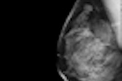
 Left: Example of a breast tomosynthesis slice holding a simulated mass. Right: Example of a breast tomosynthesis slice holding a simulated microcalcification cluster. All images courtesy of Dr. Pontus Timberg.
Image volumes can be viewed using free scroll volume browsing (FS) or in a cine loop. The standard procedure is FS, but in clinical practice observers often use a cine loop to get an overview of the images. In addition, breast tomosynthesis images may be displayed on the monitor in either vertical or horizontal orientation. The researchers evaluated the efficiency of several breast tomosynthesis image volume readings using an experimental setup. They assessed these in terms of lesion detection performance, time efficiency, visual attention, and search using jack-knife alternative free-response receiver operating characteristics (JAFROC) and eye tracking.
The researchers selected 55 normal breast tomosynthesis cases in mediolateral oblique view, which were then verified by an expert radiologist panel. The exams were acquired with a Siemens Mammomat Novation BT prototype (Siemens Healthcare). All viewing procedures consisted of FS, and three were combined with initial cine loops at three different frame rates (9, 14, and 25 fps). The presentation modes consisted of vertically and horizontally orientated image volumes.
 Example of the ViewDEX user interface showing a breast tomosynthesis slice in vertical position.
Timberg and colleagues found horizontally orientated tomosynthesis image volumes were read faster than vertical ones when using free scroll browsing only and when combined with fast cine loop. Cine loops at slow frame rates were ruled out as inefficient. Their results indicate medium frame rates are comparable to fast ones in terms of reading time, whereas breast tomosynthesis image volumes shown at slow frame rates are statistically significantly slower to read. Also, faster cine loops lead to shorter entry times.
"Studies on visual perception when reading radiology images are rather scarce," they wrote. "Most of the studies have been related to mammography, and only a few of these studies have used eye tracking."
Using eye tracking, the researchers found in horizontal presentation mode images were better aligned with the human visual field, which has wider extension horizontally than vertically. Also, peripheral vision can be used more efficiently to localize abnormalities in the tomosynthesis image volumes, in particular where lesions appear and disappear abruptly in dynamic presentations.
"Such abrupt onsets are known to capture attention in a bottom-up manner and thus attract observers' gazes," they stated. "Therefore, it should be emphasized that the real benefit lies in viewing dynamic images rather than stationary images like 2D mammograms."
The authors also noted they were surprised little time was spent in the slow cine loop, and hypothesized the observers felt uncomfortable awaiting appearances and reappearances of structures of interest, or the slow cine loop did not provide a satisfying capture of visual attention.
 Example of the ViewDEX user interface showing a breast tomosynthesis slice in horizontal position.
The relatively short time spent in the cine loop was also found in the other cine loop speeds, but this is possibly indicative of attracting visual attention more quickly, the authors added.
"Our intention is to continue our research in optimizing reading conditions of breast tomosynthesis image volumes," Timberg said in an interview with AuntMinnieEurope.com. "It would be of interest to investigate readings of other imaging modalities as we believe it is beneficial to aligning the content to the visual field, especially in dynamic presentations of 3D volumes."