Remote interpretation of imaging studies often means using a mobile device with a small screen. But virtual reality technology can overcome those limitations and offer an immersive image viewing environment without sacrificing image quality, Greek researchers reported at RSNA 2015 in Chicago.
After modifying virtual reality hardware and software to be able to support the viewing of DICOM images, a team from Evangelismos Hospital in Athens found it could produce more than 97% interobserver agreement in testing on 271 CT studies.
"Remote diagnosis of complete CT examinations performed elsewhere using our mobile virtual-reality [device] is feasible and useful," presenter Dr. Vasileios Moustakas said.
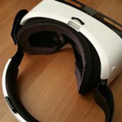
 Virtual-reality device adapted for use with imaging studies. All images courtesy of Dr. Vasileios Moustakas.
The group set out to obtain a high-resolution mobile virtual-reality prototype and to determine if it could be used to view DICOM images without compromising image quality. As a secondary purpose, the researchers wanted to verify the mobile virtual-reality system could be utilized for remote diagnosis of CT exams.
Virtual reality is a technology that creates an immersive, artificial environment for 360° around the user. The prototype developed by the Greek team provides a similar experience.
"Using our system is like standing in front of a 175-inch mega-screen, while enabling visualization at 360° around the user and having a 96° viewing angle at any time," he noted. This enables the user to move freely in all directions.
The device is completely mobile, and does not require a personal computer or additional equipment, Moustakas said. It weighs 0.3 kg (0.7 pounds) and does not require a power cord as it is powered by a smartphone with a quad-core 2.7-GHz central processing unit (CPU) and an ultrahigh-density 550 pixels-per-inch display. Both brightness and contrast were calibrated using special gray-scale patterns to match the hospital's diagnostic workstations.
Significantly, the virtual-reality device is not affected by the ambient environment.
"When you wear it, you don't see the outside world," he said. "There are no problems with lighting. Whether you are in sunlight or in a dark room, it doesn't affect the virtual-reality system. So once it's calibrated one time, it's forever; you don't have to test again."
The smartphone's 4G+/LTE connection enables DICOM images to be quickly downloaded from the server, and up to 56 DICOM images can be viewed at one time on the device, he said. Hand motions are used to scroll through images and perform functions such as zooming in and out.
 Up to 56 DICOM images can be viewed at one time on the virtual-reality device.
No effect on image quality
The researchers tested the device using 271 complete CT exams from 138 patients; some of the patients had been examined more than once and many had received multiple scans. The patients were scanned for various indications and the exams were reviewed by a consultant radiologist in the hospital. All types of CT scans (abdomen, brain, chest, lungs, and spine) and both with and without contrast were included in the research study.
Next, the CT exams were independently and remotely reviewed in a blinded fashion by another consultant radiologist using the virtual-reality system. Five critical checkpoints (such as the presence of intracranial hemorrhage, width of ventricular system, midline structure alignment, density of white matter, and middle cerebral artery density for brain imaging) were used for each of the 271 examinations, leading to a total of 1,355 total results in the study.
Using standardized reporting systems, the radiologists' reports were then evaluated to assess the image quality of the virtual-reality device in comparison with the quality of images on the hospital's workstations.
Complete interobserver agreement was found in 1,318 (97.3%) of the 1,355 results. The other 37 (2.7%) results were limited to evaluations that often have discrepancies between different examiners on the same monitor, such as assessing white-matter density in elderly patients.
"[The results show that] the use of our virtual-reality device did not affect image quality and therefore, did not alter the diagnosis," Moustakas said. "This technique can be used for remote diagnosis while avoiding the small-display limitations."
On-the-go viewing
The virtual-reality device could be used in cases where a second, more-experienced opinion is needed remotely in real-time on imaging studies. In the future, further improvements in high-resolution displays might yield even higher pixel density, leading to replacement of classic diagnostic displays with virtual-reality devices, whether they be used for mobile or nonmobile applications.
"Improvements in virtual-reality software could soon lead to substitution of classic diagnostic monitors with virtual-reality devices when visualizing 3D reconstructions," he said. "Virtual reality provides an unparalleled 3D experience."
What's more, virtual-reality devices are ideal for creating 3D models for printing on medical 3D printers, Moustakas concluded.