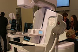
The modern radiologist works on PACS workstations in their clinical practice. So why not use a workstation-like interface to learn many of the crucial skills needed to be a radiologist? That's the theory behind Imaging Anatomy, an interactive online atlas developed by Danish radiology resident Dr. Jens Borgbjerg.
The open-access atlas is designed to assist users in developing skills for navigating volumetric datasets, said Borgbjerg of Aalborg University Hospital. Radiological anatomical structures are identified and presented in multiple planes, with an interface that functions just like a PACS workstation.
What's more, users can gain understanding of how the presentation of anatomy is affected by image manipulation tools such as zoom, pan, brightness/contrast adjustment, CT window/level selection, and caliper/angle measurement, he said.
"The idea is that such an atlas can provide familiarization with anatomy in a form that mimics the way you typically work with anatomy in modern clinical environments, and allows for assimilation of this familiarization into clinical practice," Borgbjerg told AuntMinnieEurope.com.
Need for acquiring skills
The pervasiveness of PACS enables not only radiologists but also nonradiologist clinicians to have easy access to imaging studies with vast datasets, Borgbjerg said.
"Thus, there is a variety of specialty groups needing to acquire skills in navigating/manipulating volumetric datasets and locating anatomical structures therein; these skills are prerequisites for being able to recognize abnormalities, understand radiology reports, and communicate confidently about imaging studies," he said.
While noting that several excellent radiological atlases have been developed in an attempt to assist users in developing radiological anatomical knowledge and skill, Borgbjerg said he wanted to provide an experience that was closely comparable to a PACS workstation.
Imaging Anatomy was conceived as a spinoff from a project Borgbjerg worked on as part of his residency training. In developing a Web-based application for handling the logistics of observer performance studies in imaging research, he began to experiment with creating scrollable image stacks using the HTML5 Web standard. HTML5 enables the creation of Web-based applications with desktop graphics and can be accessed through pretty much any platform -- including mobile devices -- using a modern Web browser, and without requiring third-party browser plug-ins such as Adobe Flash, he said.
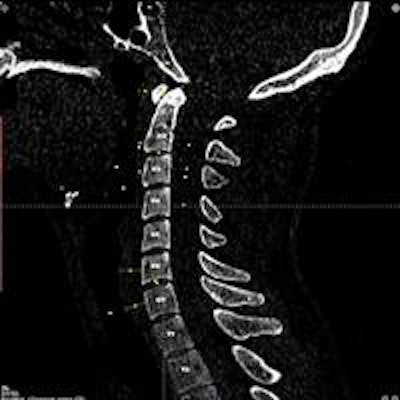
 Sagittal cervical spine CT image with anatomic landmarks on Imaging Anatomy. Image courtesy of Dr. Jens Borgbjerg.
The imaging studies found on Imaging Anatomy were acquired from Aalborg University Hospital's PACS database and were anonymized prior to use on the site, which was launched in June 2014. He shared more details on Imaging Anatomy in a poster presentation at the 2015 ECR.
Since ECR 2015, Borgbjerg has added a number of new features to Imaging Anatomy, including a module search function that will take the user directly to the relevant imaging slice, regional shading "hot spots" to better delineate anatomy, a new saving feature that allows users to save the position and size of the displayed module views, as well as general settings and a self-quiz tool for assessing anatomical knowledge.
Positive feedback
While there hasn't been any formal validation of the learning efficiency of Imaging Anatomy, Borgbjerg said he has received positive initial feedback from health professionals in various specialties at both the postgraduate and undergraduate level.
"A number of radiology residents from my own institution as well as outside have highlighted the usefulness of [Imaging Anatomy] as a resource to be utilized alongside a PACS workstation where multiple diagnostic displays are available," he said. "In this way, it is possible to reference normal radiological anatomy and thereby assist the process of recognizing structures when learning to interpret studies such as knee MRIs on a PACS."
The interactive aspects of Imaging Anatomy are also well-received. Studies have shown the new generation of medical trainees, the millennials, are tech-savvy, have an advanced readiness to use new medical technologies, and have an affinity for interactive approaches to learning, Borgbjerg said.
"Recognizing and addressing the needs of these students, while teaching basic imaging skills, is vital for successful learning outcomes," he said.
Based on the widespread use of cross-sectional imaging today and the overall feedback he's received, Borgbjerg said he believes the atlas in its current iteration should be useful to radiological trainees, senior radiologists, radiology technicians, clinicians, surgeons in multiple disciplines, and medical students.
Imaging Anatomy is being utilized around the world. So far in September, the site has been visited by users from 114 countries, he said. It has 23 anatomical modules and includes x-ray, CT, MRI, and ultrasound images.
In the future, Borgbjerg plans to incorporate additional anatomical landmarks and normal values to existing modules, and add more modules with new anatomical regions and modalities such as 7-tesla MRI scans. He also will present a new learning module at the upcoming RSNA 2015 meeting in Chicago that is designed to help users develop a search pattern for interpreting cross-sectional imaging studies.