Real-time image guidance of radiotherapy using MRI, with its high soft-tissue contrast, promises dramatically improved tumor targeting and an increase in the number of treatable disease sites. Tackling the challenge, an MR-guided cobalt-60 unit is already in clinical use and three groups are developing integrated MRI-linac systems, including a collaboration of seven Australian institutions.
In new work, the Australian collaboration, led by Paul Keall, PhD, professor at the University of Sydney and director of the Radiation Physics Laboratory, has demonstrated the feasibility of its first prototype. Constructed at the Ingham Institute in Sydney, the system combined a commercial portable linac with an ex-clinical MRI system. The prototype uses an inline configuration, where the photon beam fires parallel along the scanner bore and is therefore aligned with the B0 magnetic field. The study quantified interactions between the two systems and has paved the way for a second, more advanced system using a custom-built magnet.
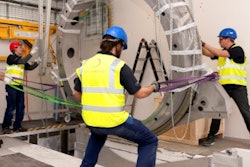
 The Australian collaboration's first prototype MRI-linac combined a portable linac with an on loan ex-clinical 1.5-tesla MRI scanner. The shielded linac (yellow) is visible on the far left. The multileaf collimator is in the middle of the photo. A copper RF screen has been removed to show the magnet.
The prototype's MRI scanner was a 1.5-tesla system on loan before disposal. It was placed in an RF-shielding cage through which the treatment beam was fired. The linac isocenter was positioned in the middle of the 60 cm diameter scanner bore. By placing the linac on a stainless steel table that, in turn, was mounted on rails, the source-isocenter was adjustable.
The researchers first assessed the impact of the linac's operation on image quality, imaging a piece of kangaroo meat and a bottle of doped water. They observed no significant effects; in images of the meat, the signal-to-noise ratio was 30 when the beam was off and 31 when it was turned on (Medical Physics, September 2016, Vol. 43:9, pp. 5168).
However, when a surface coil was deliberately placed in the beam, the background signal rose by up to 24%. The effect could be exploited, the authors wrote, and is something the collaboration plans to investigate further. "By plotting the noise in the image we can 'see' when the beam is on and maybe even relate it to the amount of dose being received by the patient," explained Gary Liney, PhD, senior MRI research physicist at the Ingham Institute for Applied Medical Research, Liverpool Hospital in New South Wales, who is the first author and the physicist leading the integration of linac and MRI hardware.
In a second investigation, the researchers assessed distortions in the scanner's B0 field as the linac was moved closer to the magnet. Phase difference maps of a sphere filled with doped water revealed increasing distortion with proximity. However, the nonuniformities were easily corrected with dynamic shimming.
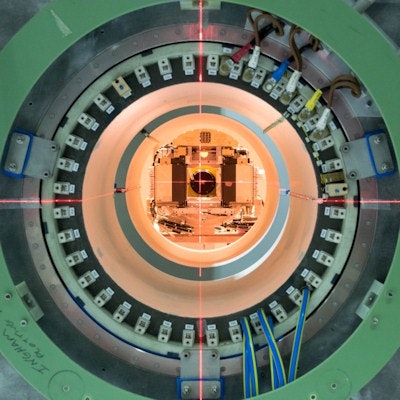
 Looking down the split bore of the collaboration's second system, currently an inline arrangement. Objects are placed in the split perpendicular to the bore and photon beam. The geometry also allows the beam to be fired down the split, perpendicular to the B0 field. A copper RF screen is placed in front of the linac during imaging and irradiation.
In a third investigation, the researchers measured interactions between the magnetic field and contaminant electrons generated by the linac beam. Irradiating film at nine points along the beam axis, they observed the electrons were focused toward it, a finding replicated by an electronic portal imaging device (EPID) behind the magnet.
Film measurements in solid water revealed an associated increase in surface dose of 300%. This effect could also be exploited clinically, wrote the authors. For instance, it could reduce the penumbral widening seen in low-density tissues like lung, reducing dose to normal tissue and enhancing dose to the target.
Working on the collaboration's second system, the researchers have since installed a custom-made 1.0-tesla split magnet, plus a gradient module and RF coil. "The next task is to try and get the software to talk to the hardware," said Liney. The imaging system -- the scanner components that generate the signal pulses and manipulate the RF and gradient coils to capture the image -- is that of a Siemens Healthineers 1.5-tesla scanner. The group must configure it to work with the lower-field magnet and other hardware that has been supplied by other vendors.
The new prototype will reproduce the inline geometry of the current system. The split design will allow phantoms to be irradiated in the space between the two magnet halves, positioned perpendicular to the MRI field and treatment beam. In the longer term, the researchers plan to investigate a perpendicular alignment of the linac and MRI system, where the photon beam is fired through the split and phantoms are aligned parallel to the bore.
© IOP Publishing Limited. Republished with permission from medicalphysicsweb, a community website covering fundamental research and emerging technologies in medical imaging and radiation therapy.