More than 10,000 7-tesla MR research examinations are already performed globally, and there are an ever-growing number of reports eloquently speaking about moving ultrahigh-field MR (UHF-MR) into the clinic. The pace of discovery is heartening and a powerful motivator to transfer the lessons learned from UHF-MR research into the clinical scenario.
These efforts are fueled by the quest for advancing the capabilities of diagnostic MRI. Momentum is gathering for multicenter trials, which will be enabled by increasingly robust and streamlined hardware and software platforms. The eye-opening quality of recent anatomical and functional images of the brain, heart, and other anatomy have created excitement in the (bio)medical and diagnostic imaging communities, and have served as a driving force for application developments.
At the same time, it is no surprise that the feasibility of 7-tesla MRI is not limited to these applications, but can also be beneficial for musculoskeletal, abdominal, breast, interventional, and last but not least, ophthalmic imaging. The implications feed into a broad spectrum of MR physics, biomedical engineering, neuroscience, neurology, radiology, cardiology, internal medicine, oncology, nephrology, ophthalmology, and other related fields of basic research and clinical science.
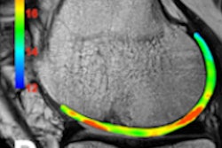
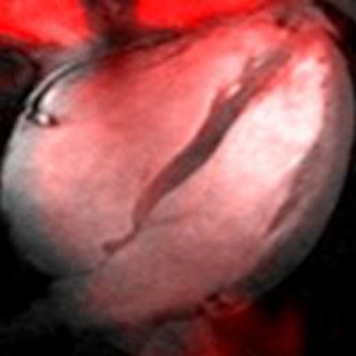
 Above: Sodium (Na-23) MR image (red) of a four-chamber view of the human heart superimposed to a 2D CINE image (gray). The Na-23 image of the heart was acquired at 7 tesla using a density-adapted short echo-time 3D radial technique. Below: Fluorine (F-19) MR image (red) of an axial view through the human knee superimposed to an anatomical image (gray). The F-19 image was acquired at 7 tesla following topical application of an ointment containing flufenamic acid -- an F-19-containing nonsteroidal anti-inflammatory drug (NSAID). A spatial resolution of (1.5 x 1.5 x 5.0) mm3 was accomplished.
Moreover, the benefits of 7-tesla innovations are already being seen at 3 tesla, where the suboptimal copy and paste approach to protocol migration from 1.5 tesla is being supplanted by the sort of application-targeted redesign that is essential at UHF. Recognizing the pace and momentum of UHF-MR, the MR vendors committed that there is no U-turn on the tesla road. In fact, they are now driving with full throttle to set up the next generation of whole-body 7-tesla systems with CE certification or U.S. Food and Drug Administration (FDA) approval eventually looming on the horizon.
One exciting development that is in the spotlight of current (pre)clinical research is that cutting-edge MR centers are exploiting UHF-MR not only for H-1 MRI but also for documenting the value of 7-tesla MRI for imaging x-nuclei such as carbon (C-13), oxygen (O-17), fluorine (F-19), sodium (Na-23), phosphorous (P-31), chlorine (Cl-35), and potassium (K-39). UHF-MR holds the promise to facilitate sodium imaging of the heart (see figure) with a spatial resolution commonly used for H-1 MR in today's clinical practice at lower fields.1
Early clinical applications of Na-23 MRI include but are not limited to the assessment of repair tissue quality following cartilage transplantation. Na-23 MRI at 7 tesla can also help to unlock questions regarding the Na+ balance and Na+ storage functions of skin,2 with the ultimate goal to provide imaging means for diagnostics and to guide treatment decisions in cardiovascular and metabolic diseases.
The added value of P-31 MRS at ultrahigh-fields was recently eloquently reported for the differentiation between nonalcoholic benign liver disease and potentially progressive steatohepatitis. Pioneering reports demonstrated the capabilities of UHF-MR for probing myocardial high energy phosphate metabolism and revealed marked superiority of cardiac P-31 spectra at 7 tesla relative to 3 tesla.
Probing phosphorylated metabolites with P-31 MR spectroscopy can be used for prostate cancer characterization at 7 tesla. The feasibility of F-19 MR of the knee (see figure) after epicutaneous application of a fluorine contained nonsteroidal anti-inflammatory drug (NSAID) was shown at 7 tesla.3 This important development contributes to the technological basis for the clinical assessment of biodistribution and bioavailability of F-19-containing medicinal compounds in vivo.
An open-minded look reveals that MR at 7 tesla is not enough. Obviously, the future of UHF-MR will not end at 7 tesla and the field is moving apace into this direction. The first pioneering reports on MR physics and radiofrequency (RF) power deposition considerations for MR at 10.5 tesla or even 14 tesla were released. The recent progress of probing the local concentrations of fluorine, sodium, potassium, and chlorine at 7 tesla provide convincing reasons for wide-bore magnets with B0 ≥ 7 tesla.
This motivation spurred the installation of a 10.5-tesla whole-body MR system suitable for cardiac and body imaging at the Center for Magnetic Resonance Research, University of Minnesota in Minneapolis, under the leadership of Dr. Kamil Ugurbil. An ongoing 11.7-tesla brain MRI initiative is spearheaded by Dr. Denis LeBihan (CEA, Neurospin, NeuroSpin, Gif/Yvette, France) and by Dr. Jeff Duyn (National Institute of Health, Bethesda, MD). Alongside this progress, there is intriguing news on potential future developments outlined in a report of the National Research Council dealing with high magnetic field science and its application.4
This report forwarded a call for a 20-tesla wide-bore MR system, a technical development inspired by the recent progress at 7 tesla as well as by the lessons learned from the design and safety evaluation of hybrid magnets running at 25-tesla.5
It is no surprise that unlocking the potential of 20 tesla is of paramount relevance for addressing spatial resolution constraints of today's MRI. In vivo MR microscopy at 20 tesla will amplify image detail. This gain will be essential for observing pathology way before the onset of clinical symptoms along with the capability of probing tissue microstructure.
Twenty-tesla MRI is conceptually appealing for the pursuit of unraveling the human brain to push the envelope at the border between (bio)physics and neuroscience. Arguably, H-1 human MR at 20 tesla might be challenging, if not elusive, in the first run. Yet, the frequencies of most x-nuclei at 20 tesla are below the H-1 resonance frequency at 7 tesla. This makes technology established for H-1 MR at 7 tesla ideal candidates to be fine-tuned for heteronuclear MR at 20 tesla.
For example, the sensitivity gain at 20 tesla is expected to reduce scan times for P-31 and Na-23 by almost an order of magnitude versus today's 7.0-tesla capabilities. While this is, for the moment, merely a vision, it promises sodium MR with a submillimeter spatial resolution in 5-10 minutes scan time, and offers the potential for probing the heart with P-31 spectroscopy in clinically acceptable scan times versus today's groundbreaking studies using scan durations of approximately two hours.
The inescapability of magnetic field strength larger than 7 tesla is highlighted by recent explorations into targeted radiofrequency heating that perhaps forms an enabling platform to advance MR thermo-theranostics.6 At 23.5 tesla, higher RF frequencies (1 GHz) result in a wavelength in brain tissue of about 4.5 cm, allowing for a more localized targeted RF heating approach versus 7 tesla.
While this might feel like utopia at first glance, a recent report underlined the potential of targeted RF heating at 1 GHz (23.5-tesla) for localized RF hyperthermia of intracranial lesions or for adjunct cancer therapy. Other applications could include targeted contrast agent, drug, or stem cell delivery afforded by local RF heating. One could even conduct a "gedankenexperiment" (thought experiment) where targeted RF heating driven by multitransmit extreme high-field MR (EHF-MR) technology is used for RF ablation versus today's invasive intracardiac catheter ablation for the pursuit of curing atrial fibrillation and other cardiac diseases.
Bringing these interventions and therapies into the clinic remains an extraordinary technical challenge, but a story worth following en route to human MRI at 20 tesla.
For more information on this area, you may want to attend the 6th Annual Scientific Symposium on Ultrahigh Field Magnetic Resonance: Clinical Needs, Research Promises, and Technical Solutions, which will be held on 26 June 2015 at the Max-Delbrueck-Center for Molecular Medicine in Berlin. The meeting has had official endorsement from the International Society for Magnetic Resonance in Medicine (ISMRM) and the European Society for MR in Medicine and Biology (ESMRMB). For free registration, please go to www.uhf-mr.de.
Dr. Thoralf Niendorf is professor and chair for Experimental Ultrahigh Field Magnetic Resonance at Charité - University Medicine Berlin and the director of the Berlin Ultrahigh Field Facility (B.U.F.F.) at the Max-Delbrueck-Center for Molecular Medicine.
References
- Niendorf T, Paul K, Oezerdem C, et al. W(h)ither human cardiac and body magnetic resonance at ultrahigh fields? Technical advances, practical considerations, applications, and clinical opportunities. NMR Biomed. 2015; DOI: 10.1002/nbm.3268 [epub ahead of publication].
- Linz P, Santoro D, Renz W, et al. Skin sodium measured with (23) Na MRI at 7.0 T. NMR Biomed. 2015;28(1):54-62.
- Yiyi J, Waiczies H, Winter L, et al. Eight channel transceiver radiofrequency coil array tailored for 1H/19F magnetic resonance of the human knee and fluorinated drugs at 7.0 tesla. NMR Biomed. In press 2015.
- Committee to Assess the Current Status and Future Direction of High Magnetic Field Science in the United States; Board on Physics and Astronomy; Division on Engineering and Physical Sciences; National Research Council. Washington, DC, USA: National Academies Press 2013:232.
- Smeibidl P, Bird MD, Ehmler H, Tennant A. New hybrid magnet system for structure research at highest magnetic fields and temperatures in the millikelvin region. Journal of Physics: Conference Series. 2012;400:052034.
- Winter L, Ozerdem C, Hoffmann W, et al. Design and evaluation of a hybrid radiofrequency applicator for magnetic resonance imaging and RF-induced hyperthermia: Electromagnetic field simulations up to 14.0 tesla and proof-of-concept at 7.0 tesla. PLoS One. 2013;8(4):e61661.
The comments and observations expressed herein do not necessarily reflect the opinions of AuntMinnieEurope.com, nor should they be construed as an endorsement or admonishment of any particular vendor, analyst, industry consultant, or consulting group.