VIENNA - Radiologists often say that the brain is the next frontier. But as diffusion MRI techniques progress, the most mysterious organ in the human body starts to unveil more and more of its secrets, and what was once inconceivable a decade ago is now almost at hand.
Researchers are now better able to understand how neurons connect with one another and how disease affects these connections in the human brain. The production and later study of maps of neural connections obtained with MRI are vital to this task. A dedicated New Horizons session covered this fascinating topic on Friday at ECR 2014.
Dr. Patric Hagmann, the chair of the session, is an attending physician and neuroradiologist at Lausanne University Hospital (CHUV, Centre hospitalier universitaire vaudois) in Switzerland. In his introduction, he described what he calls the connectome, a term he coined in his thesis on diffusion MRI and brain connectomics back in 2005.1
"We could sum up the connectome as a comprehensive map of neural connections in the brain. The production and study of connectomes is what we refer to as connectomics; it may range from a detailed map of neurons and synapses within part of, or all of, the nervous system to a description of the functional and structural connectivity between all cortical areas and subcortical structures," he said.
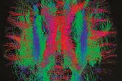
 White matter fiber pathways of the brain as depicted with MR tractography. All images courtesy of Patric Hagmann, CHUV-UNIL, Lausanne, Switzerland.
In his presentation, Hagmann introduced important concepts related to connectomics such as scaling, the relation between structural and functional connectivity, and the integration-segregation, but also showed how advances in MRI facilitate the mapping of the human connectome.
"The development of diffusion MRI and MRI-based techniques such as white matter tractography -- the computed reconstruction of images acquired during an MRI scan -- and segmentation of white and gray matter in the past decade have played a crucial role in the emergence of connectomics, by providing tools to map, in vivo, the entire human structural connectivity at a macroscopic scale," he said.
Neural fiber pathways connecting gray matter can now be represented as a network, a set of nodes and edges. To help attendees visualize this network and the effect of disease upon it, Hagmann offered the example of airline traffic.
"Areas in the brain are like airports of different sizes. You have small airports like Geneva, intermediate airports like Madrid, and hubs like Heathrow or Frankfurt. When there's a problem at a small airport, it will have a limited impact on the rest of the traffic. But when there's a problem at a hub, there will be consequences for the whole network. It's the same in the brain; some diseases affect hubs, others intermediate or small areas, and the effects on the organism will differ accordingly," he said.
A typical example of a disease primarily affecting the hubs would be Alzheimer's disease. Schizophrenia is different, with a more distributed network of alterations affecting global efficiency. "In this case, it is as if traffic were reduced by 10%; global traffic is still going, but not as well as it normally would," said Hagmann, who has been working on the topic extensively at CHUV.
Connectomics may help in the understanding of the biological basis of psychiatric disorders such as autism and schizophrenia, and Martijn P. van den Heuvel, PhD, an assistant professor in the department of psychiatry at the University Medical Centre Utrecht in the Netherlands, discussed the application of network science and the role of connectomics in his work during the session.
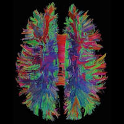
 The brain represented as a network: Image obtained from MR connectomics.
The brain has some particular network properties such as high topological efficiency, robustness, modularity, and a "rich club" of connector hubs that are advantageous for information transfer efficiency. Ed Bullmore, PhD, a professor of psychiatry from Cambridge University in the U.K., discussed how these features may be mostly explained by a drive to minimize wiring cost. Bullmore also gave an analysis of the connectome in psychiatric disorders, which may highlight the vulnerability of certain brain areas.
Using models of neural dynamics (i.e., of brain activity) is crucial to better understanding how the brain works. Gustavo Deco, PhD, a professor of information and communication technologies and director of the Center of Brain and Cognition at the Universitat Pompeu Fabra in Barcelona, Spain, has been working with Hagmann on these questions. Deco also spoke during the session about sophisticated models of spontaneous neural activity of the brain using connectome data obtained from MRI. He showed that, by comparing the model with effective human recordings, important neurophysiological details can be unraveled.
"Brain mapping models used to be very limited; the maps back then showed maybe a handful of neural connections. With the advent of diffusion MRI and tractography, we have been able to produce large scale models of at least 20,000 connections, on which we can simulate neural activity. This practice has actually become very popular," Hagmann said.
These methods are not ready to enter clinical practice yet, but MR connectomics will hopefully have a role in the diagnostic workup of the patient in the future, according to Hagmann, who suggested looking at the history of voxel-based morphometry (VBM) as an analogy. VBM is a neuroimaging analysis technique used to investigate the focal differences in brain anatomy using statistical parametric mapping. It has been mostly used to compare groups in clinical neuroimaging science. With the increasing experience in this field, VBM is slowly coming into clinical practice. For instance, the study of the atrophy of the temporal lobes on MRI is now, along with blood and neurological tests, vital in the diagnosis of Alzheimer's disease.
"Advances are being made very fast and will continue to do so," Hagmann predicted. "We are talking about things that would have been inconceivable 15 years ago, because the technology wasn't there yet."
At this pace, imaging looks set to greatly shape future management of brain diseases.
Originally published in ECR Today on 7 March 2014.
Copyright © 2014 European Society of Radiology
1 "From diffusion MRI to brain connectomics," a study based on diffusion tensor MRI data of 32 healthy volunteers and in which language networks were investigated. For more information, please visit infoscience.epfl.ch/record/33696.