UK Biobank, which calls itself the world's largest imaging study with plans to scan more than 100,000 participants, is adding imaging centers and undertaking new projects, according to the group's November 2016 newsletter.
The multimillion-pound study is currently rolling out across the U.K. with the goal of increasing awareness of various diseases, including dementia, arthritis, cancer, heart attack, and stroke. Funded by the Medical Research Council, Wellcome Trust, and the British Heart Foundation, UK Biobank currently has one imaging center near Stockport and will add others in Newcastle-upon-Tyne and Reading, the organization said.
Current projects include functional brain imaging with functional MRI (fMRI). Brain data include one kind of image for anatomy and another depicting function, revealing complex patterns of brain activity, said Dr. Stephen Smith from the Oxford University Medical Center. A third image type depicts the brain's connections.
Another branch of research is exploring fat distribution as a marker of an individual's risk of future disease, considering evidence that fat distribution, rather than the amount of fat, is key to risk assessment. Abdominal MRI offers opportunities to explore the predictive importance of particular fat deposits and relative distribution of fat, the organization said. Research involves examining fat around the liver, pancreas, skeletal muscle, kidneys, and lungs, UK Biobank stated.
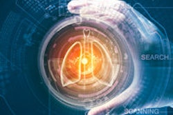
MRI is the tool being used to explore the heart chamber diameters, blood flow volumes, and changes in the heart as it pumps blood, the organization stated. Researchers are looking at the thickness of the heart wall and size, shape, and stiffness of the thoracic aorta. MRI is being used to study brain structure and function, gray-matter volumes, and the mapping of brain connections.
UK Biobank noted it has also given considerable thought to incidental findings and said that, in general, imaging is not a health check, and staff are not looking for abnormalities. When potentially serious findings are spotted during routine scan of volunteers, the findings are referred to a radiologist, who contact the patient and his or her general practitioner. About 2% of participants so far have encountered these types of potentially serious findings, of which two-thirds turn out to be not serious, the organization said.