Hip injuries are common among elite synchronized swimmers, but they form only a part of a complex clinical picture in a group of athletes that represents a major diagnostic challenge, according to researchers from a top London institution.
"The high prevalence of hip findings in asymptomatic synchronized swimmers should lead to cautious interpretation of MRI findings and astute clinical correlation," noted lead presenter and orthopedic specialist Natasa Devic at the 2015 congress of the European Society of Musculoskeletal Radiology (ESSR). "It is possible that these findings in apparently asymptomatic athletes represent tissue damage that may lead to development of future symptoms."
 Synchronized swimming is an Olympic sport that combines swimming, dance, and gymnastics. Participants perform a synchronized routine of elaborate moves, accompanied by music.
The authors' aim was to establish the prevalence of hip MRI findings in an asymptomatic group of elite synchronized swimmers. Using a Siemens Healthcare 3-tesla Skyra MRI system, they performed bilateral screening MRI exams on the 10 members of the England synchronized swimming team. All the athletes in the study group were asymptomatic.
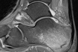
The protocol included three-plane, fat-saturated proton density, and coronal and axial proton density scans. The axial images were aligned to the long axis of the femoral neck. The images were assessed for bone, articular, tendon, and muscle abnormalities, according to Devic, who trained as a surgeon at St Thomas', St George's, and the Chelsea and Westminster hospitals in London and is currently studying for a higher degree in orthopedic biomechanics at Imperial College. She is working toward a diploma in sports and exercise medicine, and aims to pursue a career as a sports doctor.
No participants reported hip symptoms. The commonest finding in the study group was a cam lesion, found in 14 out of the 20 hips (70% of hips studied). Four out of 10 swimmers had bilateral cam lesions. The most common cam lesion morphology was the superolateral type (71%), and the anterolateral type accounted for the remaining 29%.
Six out of 20 hips (30%) had labral tears, with two patients having bilateral labral tears, and four of the labral tears had an anterior location and two were lateral. A total of 13 out of 20 hips had MRI evidence of paratenonitis (65%). Gluteus medius (70%) and iliopsoas tendons (30%) were the most commonly affected. There were no acetabular pincer-type abnormalities, and no chondral or osteochondral (OCD) lesions.
| Prevalence of MRI findings | |
| MRI finding | Prevalence |
| Femoroacetabular impingement | |
| Cam | 14/20 (65%) |
| Anterolateral | 10/14 (71%) |
| Superolateral | 4/14 (29%) |
| Bony abnormality | 0/20 |
| Pincer | 0/20 |
| Articular pathology | |
| Labral tear | 6/20 (30%) |
| OCD | 0/20 |
| Tendon | |
| Enthesitis | 0/20 |
| Bursitis | 1/20 |
| Avulsion | 0/20 |
| Paratenonitis | 13/20 (65%) |
| Gluteus medius | 9/13 (70%) |
| Iliopsoas | 4/13 (30%) |
| Muscle injury | 0/20 |
Hip pain is a common presenting complaint in athletes, and previous studies have highlighted the high prevalence of hip pathology in asymptomatic athletes, with one paper reporting a 56% prevalence of labral tears and an 18% prevalence of OCD in asymptomatic hockey players, she continued. Other groups have reported a prevalence of cam lesions in asymptomatic volunteers to be around 15%, and men are five times more likely to be affected (i.e., much lower than the prevalence of cam lesions in Devic's study).
"We can conclude that patients with these findings are more likely to be symptomatic, than the patients with cam lesions and paratenonitis," the authors wrote. "We may have seen a higher prevalence of labral tears, if we had used an arthrographic technique, as this has been shown to increase sensitivity of labral tear detection."
Follow-up studies may help determine whether these findings are a risk factor for the development of future symptoms. This was suggested in an article published last year (Khanna et al, American Journal of Sports Medicine, 30 January 2014). Khanna et al concluded that the presence of cam deformity represents a significant risk factor for development of hip pain in nonathlete volunteers, but not all patients with a cam deformity develop hip pain.