Women younger than 40 with breast cancer do not have different imaging characteristics than their older counterparts, but they do have similar genetic profiles, which have a different distribution in young women compared with the general population, French researchers have found.
Approximately 7% to 8.8% of all breast cancers are diagnosed in women younger than 40, with less than 4% occurring in women younger than 35, according to a new study published in European Radiology (6 August 2013).
Other than women with BRCA mutations (for whom screening can be beneficial), early diagnosis of breast cancer in young women still remains a challenge due to several factors:
- A very low percentage (1.9%) of women 30 to 39 years old present focal breast signs or symptoms.
- Guidelines for screening mammography in most countries exclude women in their 40s.
- Underlying dense breast tissues typically found in younger women can mask tumors, resulting in poor mammography performance.
- Younger women have different and atypical presentation of lesions compared to older women, with mammography and ultrasound more likely to be interpreted as benign.
"Little is currently known about characteristic imaging features of breast cancer in patients under 40 years old," wrote Dr. Bénédicte Bullier, from the radiology department at Institut Bergonié, Comprehensive Cancer Centre, in Bordeaux, France, and colleagues. "There have only been very few studies using analogical techniques, or with incomplete radiological data, that have reached conclusions on the frequent pseudobenign presentation of cancers in this age group."
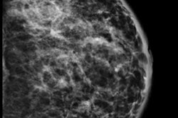
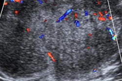
 A 34-year-old woman with 50-mm suspicious palpable lump in the left breast. Top: Mammogram (lateromedial view, spot compression). It shows no mass, no architectural distortion, just three microcalcifications. Bottom: Sonogram shows no mass but a focal heterogeneity ("nonmass"). BI-RADS 4. Biopsy under ultrasound guidance: grade III ductal invasive carcinoma HER2+. All images courtesy of Dr. Martine Boisserie-Lacroix.
In this retrospective study, the researchers sought to identify and describe the imaging characteristics of sporadic breast cancer for women younger than 40, excluding patients with proven BRCA mutations. They also sought to identify the imaging correlates of these novel molecular classes of invasive breast cancer.
The researchers reviewed radiological, clinicopathological, and biological features of sporadic breast cancers for 91 women younger than 40 from 2007 to 2012. Mammography was available for 97 lesions, ultrasound for 94, and MRI for 38.
The most common imaging features were masses; nearly all were classified as BI-RADS 4 or 5. On mammography, microcalcifications alone accounted for 31% of the lesions. There were 42.6% luminal B, 24.5% luminal A, 19.1% human epidermal growth factor receptor 2 (HER2)-enriched, and 10.6% triple-negative (TN) tumors by immunohistochemistry. HER2 overexpression was correlated with the presence of calcifications at mammography (p = 0.03), the researchers found.
TN cancers more often had an oval shape and abrupt interface at ultrasound and rim enhancement on MRI. MRI features were suspicious for all cancers and rim enhancement of a mass was a significant predictor of triple-negative tumors (p = 0.01).
There was a low incidence of ductal carcinoma in situ (DCIS, 3.1%), which is typical in a nonscreened population. The majority of cancers in the study were invasive ductal carcinoma (IDC, 92.8%) showing a high grade (48.9%, grade III).
"It is important to consider the possibility of a malignancy in symptomatic young women despite the low prevalence of breast cancer in this group," the researchers wrote. "Clinical breast examination is recommended as part of routine medical care; however, most patients in this age group present with a palpable lump that is usually found by the patient herself (78.4% in our study)."
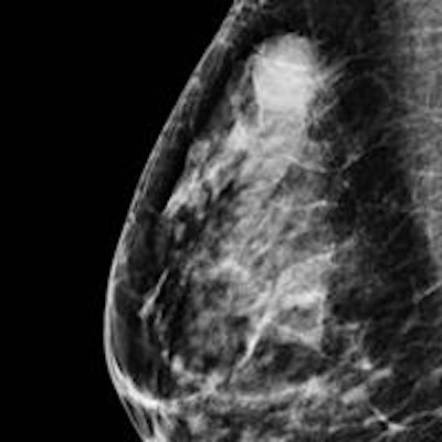
 A 33-year-old woman with 15-mm palpable lump in right breast. Top: Mammogram (mediolateral oblique view) shows an oval-shaped mass with indistinct margins. Bottom: Sonogram with shear-wave elastography shows a round mass with hypoechogenicity, abrupt interface, posterior acoustic enhancement, and a low color rating. All the benign criteria (oval mass, circumscribed margin, etc.) are not filled for fibroadenoma. This is categorized as BI-RADS 4. Biopsy under ultrasound guidance: grade III triple-negative (TN) ductal invasive carcinoma (estrogen-negative, progesterone-negative, HER2-negative cancer). TN cancers may have pseudobenign imaging features and be classified BI-RADS 3.
The usefulness of mammography to diagnose breast cancer in young women is still debatable, Bullier and colleagues wrote. Abnormal findings were visible on 84 mammograms (86.6%); the 13 false negatives involved 10 breast parenchyma density BI-RADS 3 and three density BI-RADS 4. The rate of false negatives is relatively low given the majority of dense breast compositions on mammography in their series (73.2% density type 3 and 4).
The most common abnormality on mammography was masses with or without microcalcifications (53.5%). Also, the researchers could precisely describe all the imaging characteristics and found masses were usually irregular and spiculated.
"For isolated microcalcifications, mammography is the only satisfactory investigation, and we insist on a high level of suspicion in young women with breast lumps shown to have microcalcifications, with or without a mass or density, in the region of the clinical finding," they wrote. "Finally, only two mammograms were classified as BI-RADS ACR 3. Contrary to previous studies pointing out that conventional imaging in young women is less likely to be called suspicious or malignant, we found a majority of BI-RADS 4 and 5 images."
Imaging features
Significantly more spiculated lesions were found in the luminal group, and the overexpression of the oncogene HER2 had a significant correlation with the presence of calcifications.
"[W]e think that in our study the predominance of luminal types is responsible for the high rate of irregular masses and the proportion of HER2+ cancers for the presence of microcalcifications," the researchers wrote.
As previously noted, mammography was not the only modality used. On ultrasound, the precise description of margins improved the characterization of a mass, and in this study the two masses assigned BI-RADS ACR 3 at mammography (both TN) were classified BI-RADS ACR 4 at ultrasound because of their microlobulated or indistinct margins.
Regarding the correlation between ultrasound features and the different molecular classes, the most suspicious masses were more frequently seen in luminal tumors and oval masses with microlobulated margins, abrupt interface, and posterior enhancement in TN cancers, similar to the few correlations in the literature between ultrasound features and molecular phenotypes in all age groups.
"These results indicate that imaging features in women under 40 years are not correlated with an 'age effect' as previously described and do not differ from their older counterparts," the researchers concluded.
Bullier and colleagues are planning a presentation in December about benign fibrocystic mastopathy in women younger than 40, they revealed in an email to AuntMinnieEurope.com.