Cardiac axis localization software can automatically and reliably detect the long-axis views of the heart, offering potential to decrease workload for MR technologists, a global group of researchers told delegates at the recent Society for Cardiovascular Magnetic Resonance (SCMR) meeting.
A team led by Carmel Hayes, PhD, of Siemens Research in Erlangen, Germany, found their software had strong agreement with manual localization methods, with no clinically relevant difference in more than 90% of cases during testing. Hayes presented the group's work at the San Francisco meeting.
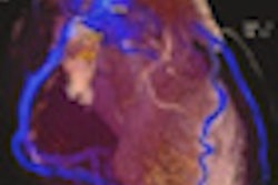
Cardiac MR typically involves acquiring standard views aligned with the heart axes, and efficient and reproducible planning requires expertise in heart geometry and anatomy, according to the Siemens Healthcare group. As a result, the team sought to evaluate the ability of their software, called AutoAlign Heart, to detect the long-axis views of the heart. They also wanted to compare, both visually and geometrically, fully automatic long-axis view planning with manual localization.
The current approach recommended by the SCMR to prescribe standard cardiac views is a multistep process that typically involves the acquisition of multiple double-oblique localizers before finally arriving at perfect long- and short-axis views in the same mode.
At the 2007 SCMR meeting, the Siemens team introduced a landmark-based localization technique, which involves the identification of five anatomical landmarks in approximate short-axis images. The long-axis image plans are then calculated based on these anatomical landmarks, Hayes explained.
"We found very good agreement between this approach and the standard classic SCMR-recommended approach," she said.
Automatic planning
Several groups, including the Siemens team, have also taken this idea one step further and investigated fully automatic heart-view planning techniques. These typically involve the acquisition of a whole-heart or whole-chest locator, and provide unsupervised heart detection, segmentation, landmark recognition, and slice prescription.
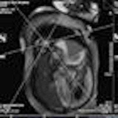
The Siemens software employs a machine learning-based algorithm that's been trained using expert-annotated images and creates a mesh representation, or model, of the heart's anatomical shape. Combining this model with landmark detection allows for calculation of slice positions and orientation.
The model was trained on 517 whole-heart MRI datasets acquired from a number of sites and a range of different scanners in patients receiving cardiac MR for a variety of reasons.
Similar to the process used in the manual planning method, the algorithm involves a coronal localizer acquisition in the first step of the process, followed by a sagittal localizer in step two and a whole-heart localizer in step three. Automatic slice positioning is performed in the final step.
','dvPres', 'clsTopBtn', 'true' );" >
The researchers tested their localization application on a Magnetom Avanto 1.5-tesla scanner (Siemens) running the vendor's syngoMR D13A software and using body matrix and spine matrix coils. Twenty-seven patients were enrolled in the study, including 17 men and 10 women. The patients were receiving cardiac MR for indications such as aortic disease (11 patients), congenital heart disease (four patients), cardiomyopathy/myocarditis (seven patients), and other reasons (five patients).
After the long-axis views of the heart were calculated and acquired in end-diastole using a single-shot TrueFISP sequence, the user then repeated the long-axis view acquisition following manual positioning of landmarks. Blinded to the acquisition approach and patient information, a radiologist then assessed the two images for visual differences and graded overall agreement using either a score of either zero (no difference), one (difference, but not clinically relevant), or two (clinically relevant difference).
Successful planning
The algorithm successfully found the heart in all 27 cases. Both manual and automatic methods yielded one major error in view planning. The software had a major error in one automatically planned three-chamber view, while the manual approach resulted in one major error for a planned two-chamber view.
The software's major error was produced in a 51-year old patient with congenital heart disease. The mistake was to be expected, however, since the software had been trained on patients with noncongenital heart disease, she said.
"So we were testing the software, in this case, in let's say, inappropriate circumstances," she said. In more than 90% of cases, there was no clinically relevant difference between the manual and automatic planning methods.
Visual agreement scores
|
Hayes also noted in the seven cases where the radiologist assigned a score of two, he actually preferred the automatically-planned views in four cases. Overall, there wasn't much of a difference in viewer preference between the methods for two-chamber views. The manual method tended to be preferred for the three-chamber view, while the automatic approach tended to be favored for the four-chamber view.
In other findings, the team found the smaller slice angulation, the smaller the visual difference between the methods.
|
"Automatic long-axis view planning is possible in the majority of clinical cases, with little or no clinically relevant difference compared with manual planning," Hayes said.